Wednesday, February 25, 2015

Mirroring Evolution


Biology concepts – bilateral symmetry, radial symmetry, planulozoa hypothesis, cephalization, last animal common ancestor, porifera, platyhelminth, cnidarian, echinodermata


Halloween was a classic slasher film. Jamie Lee
Curtis looks so young, decades before Freaky
Friday or yogurt commercials. Michael Myers
could cut a man in half with his machete, but
could he produce two mirror image halves?
Slasher movies have been around for years. The heyday of the knife-wielding madman was in the 1970’s-1980’s with films like Halloween and Texas Chainsaw Massacre. Even today we have examples, like American Horror Show, both the Asylum and the Freak Show seasons. The common theme to the movies is often someone getting something cut off or basically halved right in front of the audience.

But how many ways can you be cut in half? Top to bottom is one way, leaving you with your head attached to one half and your feet attached to the other. Or you could be cleaved through your ears and down through your body. Then you would have your nose attached to one half and your bum attached to the other.

However, there’s only one way to slice you that will give two mirror images, each with the same components. If Chucky happens to catch you through the top of your head, down through your nose and straight down to where your legs split, each half will have one eye, one arm, one leg, one ear. This can only occur because you are bilaterally symmetric. Most animals (about 99%) have bilaterally symmetric bodies, so we have to at least consider the possibility that this provides some sort of advantage.


Cnidarians like jellyfish have radial symmetry,
but not spherical. You still have to cut them in
half from top to bottom through the center. When
some move on their own, instead of floating, they
move like bilaterally symmetric animals – could
this have been the start of bilateral symmetry?
But not all animals are bilaterally symmetric, especially those that diverged earliest from the last animal common ancestor (LACA). Cnidarians (jellyfish, corals, sea anemones) are a phylum of organisms that diverged fairly early and show radial symmetry. This means that anywhere Michael Myers slashed them from top to bottom and through the center, he would always produce two mirror image halves.

If bilateral symmetry is advantageous, why are jellyfish still radial? Because it works for them; no pressure/ random mutation combination sent them on that path. Remember, evolution doesn’t have a plan, it is neither reactive nor proactive. Random mutations are always occurring, and sometimes a change in environment makes renders a random mutation advantageous. It’s simply hit or miss. If the mutation or the pressure occurred at some other time, they would miss each other.

Radially symmetric animals tend to be sessile (non-moving), free-floating, or very slow movers. They don’t chase prey down, so they don’t need to be fast. This is the advantage of bilateral symmetry; it coordinates movements so that an animal can move in a particular direction faster. In fact, one 2102 paper puts forth the idea that maneuverability is the main reason for the maintenance of bilateral symmetry in animals.

However, fast movement wouldn’t be much use if you didn’t know where you were going. This is why bilateral animals also have a head. A head is a place to store your sensory apparatus and your neural tissue to process those sensory inputs. You think its an accident that our brain is located the same place as our eyes, ears, nose, and mouth?

Slow or sessile animals (like cnidarians) that filter feed or catch what runs into them have no head. They have few sensory neurons, and only loosely associated ganglia of neural tissues spread throughout their bodies.


Humans decided to become bipedal (two footed)
and this made it hard for us to lead
the way with our head. Our foramen magnum
(hole where spine emerges) moved down and
below our skull to support the weight of our head
and keep our sensory organs pointed in
the right direction.
But if you’re going to have a head to sense the environment and help you move well in one direction – where should you put it? At the front of course. Bilateral animals also evolved to have an anterior and posterior end – the anterior end being the direction that they move. And this is where we find their head.

Bilateral animals have a head, and radial animals don’t have a head. This sounds like a fairly plain story – as animals diverged and evolved, some developed a head and became bilateral. Or..... did they become bilateral and then develop a head? Maybe the animals can tell us which way it was.

The flatworms (platyhelminthes) were the first divergence of animals to have their neural ganglia clustered in their anterior end. Going along with this, they have sensory systems located at that end too. They have eyespots, although they are really just patches that detect light or dark.


Platyhelminthes have a define head ganglion of
nerves and have started develop more senses at
the anterior end; see the sensors that stick up.
And they move faster and in a straight line. They
are headed, and headed in a particular direction.
Platyhelminthes have mechanosensors to know if they touch something, and they have chemical sensors to sample the water in front of them. That sounds a lot like our eyes, mouth, nose, and sense of touch. Since these are all at that anterior end, I call that a head. The worms are longer than they are wide, and they move primarily in one direction letting their head lead the way.

So we have gone from animals with no head and radial symmetry to animals with a head and bilateral symmetry. This doesn’t help answer the question of which came first. Aren't there any animals in between?

Yes, there are, and they give us a little bit of a clue as to which came first. The ctenophora (pronounced "ten", cteno = comb and phora = bearing) is a phylum of animals that lie between the cnidarians and the platyhelminthes. Ctenophoran animals are the comb jellies. Both cnidarians and comb jellies have been around for over 500 million years, so they’ve had time to settle in to a niche.

The comb jellies look round at first glance, but their architecture is a bit more complex than the cnidarian jellyfish. They have internal and external features that allow only for two planes of symmetry that give mirror images (see picture). These especially include the combs, rows of fused cilia that line their sides, and the fact that they don’t have stinging cells (cnidocytes). Remember that ONLY cnidarians have cnidocytes.


Here is a cladogram that shows the divergence of
each phylum of animal from their last common
ancestor. Ctenophores and cnidarians diverged from
each other recently (or did they, see article). Starfish
diverged after everyone else on that end was
bilateral, yet they are radial as adults. What gives?
Just as the ctenophora lie between the radial cnidarians and the bilateral flatworms, their symmetry lies in the middle as well. Two planes (bi) in an otherwise radial animal = biradial symmetry.

A 2004 study investigated the relationships between biradial and bilateral animals in evolution. If biradial is the link between radial and bilateral, then would seem to suggest that bilateralism occurred before cephalization.  Called the Planulozoa Hypothesis, the authors suggests that ctenophora are the sister clade of bilateralians, and that all three of the groups – cnidarians, ctenophora and bilaterals – are the descendents of a single bilateral ancestor.

Ctenophora larvae have bilateral features, so this supports the planulozoa hypothesis (the free swimming larvae of all three phyla are called planulae). This would then suggest that cnidarians were once bilateral and then returned to radial symmetry.

Additionally, the if the planulozoa hypothesis holds, then bilateralism would seem to predate cephalization (development of a head). The larvae of ctenophores and some ctenophore features show that a move to true bilateral symmetry came before platyhelminthes and the emergence of a head. The conclusion – the streamlined body came before the head. But that confuses me, one isn’t much good without the other.
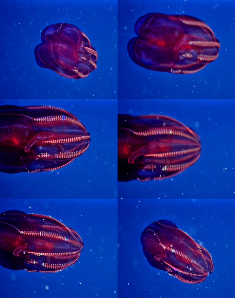

Ctenophores – the comb jellies, often show
bioluminescence. They only have two perpendicular
planes of mirror image symmetry. You can see the
fused cilia that form the combs on each ridge.
Wait a minute, there’s a fly in the bouillabaisse. Ctenophores have a nervous system that is more complex than many other animals – it’s just not centralized to a head. Centralizing the nervous system, with the sensory processing and muscular control, is a crucial part of cephalization. They seem to developed a strong neural system without adding the head itself.

A 2014 study of the genome of several ctenophores showed that they do not have the same neuron-building gene regulation pathways as any other phylum of animals, and they only use one of the most common neurotransmitters; all their other neuron signalling molecules are unique to ctenophores alone. This suggests that they evolved radically differently than the phylums around them, cnidarians and flatworms. This does not support the planulozoa hypothesis at all. Ctenophores may have developed all on their own and therefore can't help us answer the question of which cam first the bilateral body or the head.

Other things about symmetry development make you say, “Huh?” as well. Look at that same cladogram of animals above – see the right side where the sea star is located? What’s a radially symmetric animal doing way over there after everyone else switched to bilateral symmetry?

The echinodermata (sea stars, brittle stars, sea cucumbers; echino = spiny, and derm = skin) also support the planulozoa hypothesis, since they seem to have undergone the same regression as the cnidarians. Echinoderms include the brittle stars, sea stars, sea cucumbers, barnacles and sea urchins. They have bilateral symmetry as larvae, but many of them become radial (pentaradial or such, depending on the number of arms) when they become adults.

Secondary radial symmetry is term for when a bilateral larva becomes a radial adult; but it is more interesting than that. The easy way for that transformation to occur would be for the arms to grow out of the larva, with the top (aboral) and mouth (oral) sides remaining the same. But that’s not how it happens.


The brittle star, an echinoderm, walks like bilateral
animal, even though it assumes pentaradial symmetry
as an adult. One arm acts as the head, and two arms
on each side work as mirror images. When it wants to
turn, it just assigns another arm to be the head.
The swimming larva becomes sessile by attaching itself to something on the sea floor. Then one mirror image side (right or left) becomes the oral side, while the other half become the aboral side of the adult. To do this, all the arms must grow from one half, and many tissues and organs are actually lost from the larva when it becomes an adult. This seems like a lot of work just to go backward in evolution.

But like I say, it works for them. The adult sea stars and other echinoderms are fairly slow. Their lifestyle doesn’t require a head or a bilateral body, so they went biologically simpler and energetically cheaper and returned to radial symmetry. All the mechanics were still in their genomes - it was really pretty smart.

However much they have tried to regress as adults, the brittle stars seemed to have retained at least a little bilateral activity. The way they move is a lot like a bilateral animal, according to a 2012 study. One arm points forward, the direction they are traveling. The arms on either side then push the animal along, like a crawling bilateral animal. I guess you can’t completely go home again.

Next week – Bilateral animals are simple - just two mirror images, right? Well no. You won’t believe the number of complex animals that break symmetry in order to give them a unique shape or function.


Moroz, L., Kocot, K., Citarella, M., Dosung, S., Norekian, T., Povolotskaya, I., Grigorenko, A., Dailey, C., Berezikov, E., Buckley, K., Ptitsyn, A., Reshetov, D., Mukherjee, K., Moroz, T., Bobkova, Y., Yu, F., Kapitonov, V., Jurka, J., Bobkov, Y., Swore, J., Girardo, D., Fodor, A., Gusev, F., Sanford, R., Bruders, R., Kittler, E., Mills, C., Rast, J., Derelle, R., Solovyev, V., Kondrashov, F., Swalla, B., Sweedler, J., Rogaev, E., Halanych, K., & Kohn, A. (2014). The ctenophore genome and the evolutionary origins of neural systems Nature, 510 (7503), 109-114 DOI: 10.1038/nature13400

Holló, G., & Novák, M. (2012). The manoeuvrability hypothesis to explain the maintenance of bilateral symmetry in animal evolution Biology Direct, 7 (1) DOI: 10.1186/1745-6150-7-22

Wallberg, A., Thollesson, M., Farris, J., & Jondelius, U. (2004). The phylogenetic position of the comb jellies (Ctenophora) and the importance of taxonomic sampling Cladistics, 20 (6), 558-578 DOI: 10.1111/j.1096-0031.2004.00041.x

Astley HC (2012). Getting around when you're round: quantitative analysis of the locomotion of the blunt-spined brittle star, Ophiocoma echinata. The Journal of experimental biology, 215 (Pt 11), 1923-9 PMID: 22573771


For more information or classroom activities, see:

Biologic symmetry –

Ctenophora vs .cnidarians –

Echinoderms -


Wednesday, February 18, 2015

Space – It’ll Mess You Up


Biology concepts –  undulipodia, primary cilia, motile cilia, ependyma, spaceflight, pathology, osteopenia, radiation damage, osteoblast/osteoclast, osteocytes,


No one wanted the elation of the moon visit to
turn to disaster as a moon germ spread through-
out the world and killed every living thing. So
they moved the astronauts from splashdown to
airstream. What a bummer that would have
been to go all Andromeda Strain…. although it
might have saved us from Watergate.
Going into space is an engineering triumph, but it isn’t without its biologic difficulties. When the Apollo 11 astronauts returned from the first visit to the moon, Richard Nixon had to congratulate them through the window of their mobile quarantine van.

Exposing humans to space germs would be bad, but the problems go the other way too. Astronauts have to deal with many changes to their body due to the reductions in sunlight, exercise, circadian rhythm, and perhaps most of all – gravity. You’d be surprised how much microgravity (in space there is still a little gravity) can mess with your body – and alot of it has to do with some of the smallest parts of your cells – the primary cilia.

In fact, a 2004 study showed changes in human gene expression during regular gravitational field changes due to sun and moon right here on Earth. If the small changes in Earth's gravity brought about by the changing position of the Sun and Moon can have measureable effects, imagine how big a deal going into space must be. A 2008 study on Rohon-Beard cells (developmental neurons in fish and amphibians) showed that they lost primary cilia when in microgravity. So gravity matters, and it matters in part because of what it does to primary cilia.

Life on Earth has evolved in gravity. Our bodies have come to expect a pull to the center of the Earth and for all the air of the atmosphere above them to be pressing down on them. These forces must have molded our anatomy and biochemistry to some degree. So if you take us off the Earth, shouldn’t we expect some problems?

Microgravity alters the responses of the body. Processes are lost and balances are shifted. Problems caused by these changes can manifest in two ways; 1) they could cause problems while in space, or 2) they could cause problems when the astronauts return to normal gravity.


Even though the uniforms look Russian, this
footage comes from the US Air Force film
archive. They were in a C-131 doing parabolic
arcs that would provide 15 seconds of
weightlessness. The idea was to see if cats in
space could land on their feet. It turns out they
can only do it if they know which way is down.
I think the ASPCA might have a thing or two to
say about this.
So what are some of the problems associated with long term spaceflight, and how might your primary, immotile, cilia be involved? While some might be considered pathologies, others are merely adjustments that the body makes according to its own regulatory systems. Sometimes, those adjustments then produce unwanted results.

Muscles and Blood Cells
Reduced gravity means that certain parts of the body don’t get stressed. Muscles pull against resistance; no resistance means no work for the muscles. Think about pushing off the walls of the International Space Station (ISS) while floating around. The necessary force is greatly reduced, so the muscle doesn’t get worked. When muscles don’t get exercise they atrophy (a = no, and trophy = food), like the legs of paraplegics.


A 2010 study confirmed that spaceflight has an affect on muscle fibers. The soleus muscle of the back of the leg lost 20% of its fibers over a 180 day mission on the ISS. Peak force was lower by 35%. The types of fibers were different as well, switching mostly to weaker thin fibers. Exercise made a little difference, but if the astronauts were asked to move a bed soon after landing, they’d have to call Two Guys and a Truck.
Your heart is a muscle; the heart does less work in space too, mostly because it can pump easier and still move the same volume of blood. We can’t say that primary cilia aren’t involved in these muscular and cardiovascular adaptations, we just don’t have evidence for it yet.

In similar fashion, it’s possible that primary cilia aren’t involved in the decreased red blood cell production during spaceflight. A 2000 study shows that blood volume is decreased in space, and this triggers lysis (popping) of the youngest red blood cells (neocytolysis).

On the other hand, immune cell function (white blood cells) rather than number is decreased in space. Astronauts are very prone to infections when they return to Earth, and sometimes even in space. A 2012 study showed that a significant percentage of astronauts manifest viral, bacterial, and fungal infections.


The immune synapse is mediated by the
presenting of an antigen by one cell to activate
another cell. Many receptors and co-receptors
are involved, and apparently putting them all in
the right place requires IFT proteins. The bottom
image shows the large synapse between a
dendritic cell (blue) and  a T cell (yellow).
Defects in blood cell function in space lead us to a big exception. Blood cells are some of the few cells that don’t have primary cilia. It probably has something to do with the fact that they circulate; their shape is important for their movement through vessels.

However, immune cells still express IFT proteins. We learned previously that IFT proteins mediate the building and the function of both motile and immotile cilia. A 2011 study suggests that the gap between the immune cell that presents a foreign body (antigen presenting cell) and the immune cell that will react to it (often a T lymphocyte), is controlled by IFT proteins. The hypothesis is that this immune synapse (synapse is Greek for join together) is the functional equivalent of the primary cilium for immune cells. Primary cilia are important even when they aren’t there.

Bones
Extended spaceflight wreaks havoc on your skeleton. The reason is that your bones need gravity to keep them growing. Yep, your bones are always growing, ….of course they’re always breaking down too.

Bones are dynamic. Osteoblasts (osteo = bone, and blast = germ) build new mineralized bone, while osteoclasts (clast = breaker) consume bone. As a result, you basically have a completely new skeleton every seven years, every two years if you're a child. Why is this necessary? Because bones are constantly responding to changes.

Consider weight lifting - the pulling of muscles on the bones to which they are attached stimulates osteoblast and osteoclast activity. Growing muscles are bigger, which means they need bigger attachments to bone. To accommodate this growth, the bones have to remodel themselves; more bone here, less there.


The left micrograph shows the difference between
osteocytes and osteoblasts. The –cytes are in the
lacunae while the –blasts are on the edge of the
matrix. The right image shows a lacuna and the
canaliculi connected to it. The osteocyte with its
primary cilium is in the lacuna. Mechanical loading
moves the fluid in the canaliculi and bends the cilium.
The growth of bone is mediated by the osteoblasts, which exude a dense form of collagen and some bone-specific proteins. This matrix becomes impregnated with calcium to become mineralized bone. Osteoblasts become trapped inside the matrix they lay down, surrounded by a small fluid-filled cavity called a lacuna. All the lacunae are connected by long fluid-filled tubes called canaliculi. This creates a huge network of osteocytes (ie. what osteoblasts are called when they are trapped in a lacuna).

Gravity and other forms of mechanical loading (putting weight or pressure on the bone) affects these osteocytes. Just walking on Earth is a source of mechanical loading on bone. The force produces a signal from the osteocytes to the osteoblasts to lay down more bone. A 2104 study has developed a model system to define how osteocytes signal osteoblasts, but we already know some things.

Like squeezing one of those worry dolls whose eyes bug out, mechanical stress on the lacunae brings movement of the fluid in the canaliculi and the lacunar network. This bends the primary cilia of the osteocytes trapped in the lacunae and triggers the signal that the bone is under a load.

The response to loading is a release hormones and other molecules that both stimulate osteoblast activity and call for the differentiation of bone stem cells to become osteoblasts. Osteoclasts are sill doing their job of dissolving bone matrix, but the stimulation of osteoblasts in the loaded area tips the balance to more bone formation in that area.


NASA and other space agencies try to fend off some
of the space changes in physiology by having the
astronauts exercise in space. It doesn’t work for
muscle, and not much for bone. Usually it works
better if they strap the astronauts down to simulate
some gravity.
In space, there is much less loading of the bones. Walking doesn’t have gravity to deal with, and muscles have to work so little because there is little pushing against them. The net result is that there is less shear stress in the bone lacunar network and therefore less bending of the primary cilia. Therefore, the pendulum swings toward osteoclast activity and results in osteopenia (penia = poverty).

The astronauts don’t have osteoporosis. Osteopenia and osteoprosis are two different things. Osteopenia means less bone production, but the bone that is produced is densely mineralized. Osteoporosis results from adequate bone formation but poor mineralization of that bone.

Osteoporosis is caused by many other things, and can be bad – but not as bad as space osteopenia. An astronaut can lose 10x as much bone as an osteoporosis patient in just a six-month stay in space. You go into space as a virile astronaut and come back as frail as your great grandmother.

Balance
We learned last week about the role of primary cilia in the vestibular system (balance). The semicircular canals in our inner ear track rotation in space, but the utricle and saccule sense gravity and acceleration in a straight line. These otlithic organs use otoconia - little mineralized bits on the ends of sterocilia on hair cells to detect movement and gravity.


The semicircular canals are for rotational changes
and have the cupula to mediate movement. The
utricle and saccule are for gravity responses. The
macula of each is the region where the otoconia
mediate the movement of the hairs in response to
changes in their position relative to gravity.
Two recent studies suggest that primary cilia mediate balance problems in astronauts. First, a 2014 study shows that otolithic organ function is reduced in patients with primary cilia diseases and that at least one reason for this is that they have malformations of the otoconia. Second, microgravity leads to larger than normal otoconia and affects primary cilia function according to a 2000 study. For astronauts, this would be a reactive adaptation, becoming a problem only when normal gravity is re-established. The adaptation is O.K. as long as you remain in space forever. Problem solved.

Radiation
Ionizing radiation is a big problem in space. The atmosphere and ozone layer that protect Earth life from much of the radiation that could damage our DNA. Besides creating mutations in DNA, the radiation of space can mess with primary cilia.

Usually solitary structures on vertebrate cells, ionizing radiation can change primary cilia number. A 2012 study showed that after radiation exposure, some cells had multiple primary cilia. These all came from the same ciliary pocket, and were probably due to aberrant basal body formation. Surely this is going to mess with any affected cell’s function, considering how important primary cilia are for sensing the cell’s immediate environment.

Next week, we should take a look at ourselves in the mirror. Animals may look symmetric, but it's more complicated. Do we have a head to lead our body, or did our evolution of our body give us a reason to have a head?


Finetti, F., Paccani, S., Rosenbaum, J., & Baldari, C. (2011). Intraflagellar transport: a new player at the immune synapse Trends in Immunology, 32 (4), 139-145 DOI: 10.1016/j.it.2011.02.001

Conroy, P., Saladino, C., Dantas, T., Lalor, P., Dockery, P., & Morrison, C. (2014). C-NAP1 and rootletin restrain DNA damage-induced centriole splitting and facilitate ciliogenesis Cell Cycle, 11 (20), 3769-3778 DOI: 10.4161/cc.21986

Troshichev, O., Gorshkov, E., Shapovalov, S., Sokolovskii, V., Ivanov, V., & Vorobeitchikov, V. (2004). Variations of the gravitational field as a motive power for rhythmics of biochemical processes Advances in Space Research, 34 (7), 1619-1624 DOI: 10.1016/j.asr.2004.02.013

Fitts, R., Trappe, S., Costill, D., Gallagher, P., Creer, A., Colloton, P., Peters, J., Romatowski, J., Bain, J., & Riley, D. (2010). Prolonged space flight-induced alterations in the structure and function of human skeletal muscle fibres The Journal of Physiology, 588 (18), 3567-3592 DOI: 10.1113/jphysiol.2010.188508

Rimmer J, Patel M, Agarwal K, Hogg C, Arshad Q, & Harcourt J (2014). Peripheral Vestibular Dysfunction in Patients with Primary Ciliary Dyskinesia: Abnormal Otoconial Development? Otology & neurotology : official publication of the American Otological Society, American Neurotology Society [and] European Academy of Otology and Neurotology PMID: 25226371


For more information or classroom activities, see:

Bone –

Space travel and the body –

Vestibular sense -

Wednesday, February 11, 2015

Thinking Skinny Thoughts Won’t Help


Biology concepts – undulipodia, primary cilia, chemosensing, obesity, depression, hydrocephalus, lithium

Winston Churchill once said that men occasionally stumble on the truth, but most people pick themselves up and carry on as if nothing had happened.


Gregor Mendel was Augustinian monk who really
joined the order because they would allow him to
study and learn for the rest of his life. Sounds like
the gig I would enjoy. Since he was a monk, do you
think he got angry that his discoveries were
ignored for 35 years?
In some cases we are shown the truth but don’t recognize it, as with Gregor Mendel’s discovery of the laws of genetics. Using his various pea plants, the Augustinian friar’s work was presented in 1865 and published 1866 – and then was forgotten for decades.

Mendel's paper was referenced only three times over next 35 years and his work wasn’t rediscovered until 1900. Two scientists gave Mendel much credit for the primacy of his work, but it really wasn’t until a fourth individual, William Bateson, came along that Mendel became widely known and his work accepted. It was Bateson coined the phrase “Mendel’s laws of inheritance.” Why did he champion Mendel so greatly – because he questioned Darwinism as incomplete. Well, it was incomplete at that time.

Just as the world was rediscovering Mendel, the primary cilium was discovered for the first time. The world didn’t exactly ignore it; we just had to wait for technology to catch up. Zimmerman first described the solitary hair sticking out of most cells in an 1898 German paper, but the next significant paper discussing primary cilia didn’t appear until 1961! We had to wait for the electron microscope and molecular biology to catch up.


The electron microscope was the piece of
equipment that allowed for deeper investigation
of the primary cilium structure. Even though the
first electron ‘scope was operational in 1931 (M.
Knoll and E. Ruska, inventors), it still took 30 years
to turn it’s power on the primary cilium. Was it
considered just an immotile cilium, or did someone
suspect it had more jobs to do? Molecular biological
techniques in the 1990’s answered that question and
led to an explosion of study on the solitary antenna
of most cells.
Last week we discussed how primary cilia are like the antennae of cells, they stick out into the extracellular environment and react to flow pressure (kidney tubule cells), vibration (hair cells of cochlea), chemicals (hormones and such) or even light (photoreceptor cilia). Now we’re realizing some of the amazing things that this sensing controls – your brain for instance.

You know that your brain works by transmitting electrical impulses through specific neural pathways. But chemistry in the brain is just as important as electricity. Hormones, neurotransmitters, even non-chemical signals like temperature and flow are converted to chemical and electrical signals via primary cilia.

As with so many other things, we learn biology best by studying what happens when things go wrong. You won’t believe the diseases that are being linked to this most innocuous of cell structures. Without any exaggeration, primary cilia make you smart, skinny, and happy. Let’s find out how.

Inside your brain are fluid filled cavities called ventricles. The ventricular system of the brain is connected through the ventricles and travel part way down your spinal column as well. They are filled with cerebrospinal fluid (CSF) and this fluid has many functions.


Here is the ventricular system of the vertebrate
brain. Blue = lateral ventricles, cyan= inter-
ventricular foramina, yellow = third ventricle,
red = cerebral aquaduct, purple = fourth ventricle,
green = central canal. What the image doesn’t show
is the connection that allows CSF to surround the
brain in the subarachanoid space, between the
brain and the skull. This is where 85% of the
CSF can be found.
Like an internal helmet, one of the functions of the CSF is to cushion the brain, acting as a shock absorber. But it does so much more than that. CSF also stabilizes the chemistry of the brain, and helps with blood perfusion by mediating the pressure in the cranium. Finally, the CSF removes waste products from the central nervous system.

The cells that line the ventricles are neuroepithelial cells called ependymal cells. They have motile cilia (2˚ cilia), as well as microvilli – which we learned last week aren’t cilia-like at all. The ependymal cell cilia beat in a specific direction depending on where they are in the system. The coordinated beating keeps the CSF flowing through the ventricles; flow is key to its functions.

The microvilli have a different job; they absorb CSF and transfer it into the brain tissue as a way of keeping the brain in the proper chemical environment. In this way, it helps mediate the CSF functions described above.

But there is a second cell type in the ependymal layer. B1 cells are pre-ependymal cells. When called upon, they differentiate to form more ependymal cells. These B1 cells are located just below the ependymal layer, but they have small areas where they stick up and touch the CSF. And here they each have a primary cilium.

A 2014 study showed that the B1 primary cilia actually control the function of the ependymal cell motile cilia. And since the motile cilia of B1 cells control pressure and flow of CSF in the ventricular system, it’s really the primary cilia who are in charge.

Because of this, a problem with the motile cilia of the ependymal cells or the primary cilia of B1 cells leads to disrupted CSF control and hydrocephalus. Hydrocephalus (hydro = water, and cephalo = brain) leads to increased intracranial pressure and this is lethal for neural tissue. Mental retardation, other complications, and death are the results of hydrocephalus.

So we already see that these short projections that were ignored for so long have one crucial job in the brain. But there’s more. It isn’t just their presence that matters; it’s their length.


Huntington’s chorea, or just plain Huntington’s
disease, is insidious; it’s lethal and there is no
treatment. It results in debilitating movements of
the motor system. Even though the genetic mutation
is with you your whole life, the disease doesn’t show
up until middle age, probably after you have had kids.
So you don’t know that you passed it on until it’s too
late. There is a test for it – would you want to know
if you had it?
Primary cilia have specific lengths in different cell types. Too long or too short and it’s like they aren’t there at all. In kidney tubule cells, increased urine flow bends the cilia, so they transmit signals to the cells, but too much signaling would be bad. Increased flow shortens the primary cilia so they become less responsive, and this is the control mechanism. In the ependymal layer, both motile and primary cilia length are crucial.

A genetic problem in a single cilial gene leads to a disease called Huntington’s chorea (means dance for the strange movements the patients make). A 2011 paper showed that the mutation lengthens both motile and primary cilia in the ventricles.  This in turn alters the beating of the motile cilia and disrupts flow of the CSF. This isn’t the only defect in the disease, but changes in CSF are thought to exacerbate the disease.

Interestingly, the changes in intracranial pressure via primary cilia changes can lead to obesity. How could CSF and eating be connected? Well, in a couple of ways – let’s investigate further.

There are several syndromes that include obesity in their list of symptoms, diseases like Bardet-Biedl syndrome, Carpenter syndrome, and others. The commonality in these diseases is that there are mutations that affect some aspect of primary cilia function, production, or maintenance. Changes in primary cilia can affect your weight?


No big message here, just thought the primary cilium
looked like Alfalfa from Our Gang. Length is crucial for
primary cilia– I suppose Alfalfa kept his a particularly
length too.
A 2007 paper narrowed down the subset of cells where the primary cilia are disrupted by putting different primary cilia under the control of different regulators in mice. Then they could wait until the mice grew up and turn off the primary cilia in various cell types. They found that it is just the POMC neurons in the hypothalamus that regulate obesity. This means that it is a brain and behavioral issue, not a problem with energy metabolism in the body.

POMC neurons make alpha-MSH and multifunctional hormone. This is released from the POMC neurons and acts on downstream pathways to tell you to stop eating. If the primary cilia on the POMC neurons are too short or absent, you experience hyperphagia (hyper = beyond, and phagia = eating), ie. compulsive eating. You just can’t stop eating.

Many of the syndromes that start as primary cilia problems show both compulsive eating AND hydrocephalus. So this explains how hydrocephalus may affect obesity in one, way, but there’s another. If you have a brain injury that damages the ependymal or B1 cilia, then hydrocephalus might result. The POMC neurons are located right next to the third ventricle, so increased intracranial pressure during hydrocephalus can damage them and lead to compulsive eating directly.


Notice how close the third ventricle is to the POMC
neurons of the hypothalamus. Hydrocephalus alone can
induce changes in primary cilia length on them so they
won’t respond to insulin or leptin. Then you
eat compulsively.
The question remains as to what signal(s) the POMC primary cilia are sensing in order to tell you to stop eating. It is probably several, chemicals that say you are full or have enough fat. Leptin, the hormone released by fat cells is certainly one of them. A 2014 study in obese mice with leptin deficiency or leptin resistance proved this. The primary cilia on POMC neurons in hypothalamus are short in these mice due to lack of leptin signaling, and therefore they don’t work well and don’t stimulate alpha-MSH release.

So, here we have a miniscule part of your neurons cells that, if not exactly the needed length, can take away your intelligence AND your beach-ready physique. But it gets worse.

Many of these same ciliopathies (disease of cilia function) are also associated with clinical depression. The problem is, we don’t know how they lead to depression. Depression is often thought to be a problem of serotonin signaling in the brain, but it can be multifactorial. Here’s one interesting result though – lithium lengthens primary cilia.

Lithium is used to treat depression, and we don’t yet really know why it works. But lithium also increases the length of primary cilia in many cell types of the brain. Considering that many depressed people gain weight, could depression and compulsive eating be linked by primary cilia length? Lithium treats them both. This could explain why people coming out of depressive episodes often lose weight.

The popular soft drink &-Up contained lithium citrate
until 1950. It is used as a mood stabilizer now, and we
know it promotes weight gain, but here they advertize it
as slenderizing. The name, 7-Up, is a mystery, but the
atomic mass of lithium is seven - hmmm.

Sounds like we’re really on to something here. People who are treated for depression and get better often lose weight. Is it because they a) feel better and then do more activity, or is it because 2) their POMC primary cilia are longer and this suppresses their appetite?

No way for number two. Biology is never that simple. It turns out that one of the major side effects of lithium treatment for mood stabilization is weight gain. It has to do with lithium affecting the function of the thyroid gland, this being one of the major regulators of your metabolism. Your metabolism slows down and you gain weight.

Maybe if we just inject the lithium into the brain ventricles…. You want to volunteer for that weight loss program?

Next week, how can primary cilia control whether mankind ever gets to step foot on Mars or help the Enterprise on its five year mission to seek out new worlds? By controlling bones….. no, not Bones McCoy, just bones.


Tong, C., Han, Y., Shah, J., Obernier, K., Guinto, C., & Alvarez-Buylla, A. (2014). Primary cilia are required in a unique subpopulation of neural progenitors Proceedings of the National Academy of Sciences, 111 (34), 12438-12443 DOI: 10.1073/pnas.1321425111

Han, Y., Kang, G., Byun, K., Ko, H., Kim, J., Shin, M., Kim, H., Gil, S., Yu, J., Lee, B., & Kim, M. (2014). Leptin-promoted cilia assembly is critical for normal energy balance Journal of Clinical Investigation, 124 (5), 2193-2197 DOI: 10.1172/JCI69395

Davenport JR, Watts AJ, Roper VC, Croyle MJ, van Groen T, Wyss JM, Nagy TR, Kesterson RA, & Yoder BK (2007). Disruption of intraflagellar transport in adult mice leads to obesity and slow-onset cystic kidney disease. Current biology : CB, 17 (18), 1586-94 PMID: 17825558

Keryer, G., Pineda, J., Liot, G., Kim, J., Dietrich, P., Benstaali, C., Smith, K., Cordelières, F., Spassky, N., Ferrante, R., Dragatsis, I., & Saudou, F. (2011). Ciliogenesis is regulated by a huntingtin-HAP1-PCM1 pathway and is altered in Huntington disease Journal of Clinical Investigation, 121 (11), 4372-4382 DOI: 10.1172/JCI57552

Miyoshi, K., Kasahara, K., Miyazaki, I., & Asanuma, M. (2009). Lithium treatment elongates primary cilia in the mouse brain and in cultured cells Biochemical and Biophysical Research Communications, 388 (4), 757-762 DOI: 10.1016/j.bbrc.2009.08.099


For more information or classroom activities, see:

Ventricular System –

Huntington’s disease –

POMC –

Lithium -