Showing posts with label endosymbiosis. Show all posts

Thursday, December 28, 2017

When Is A Chloroplast Not A Chloroplast?

Biology concepts – gravitropism, plastid, chloroplast, chromoplast, amyloplast, leucoplast, malaria parasite

Believe it or not, the way plant roots know to grow into the dirt is related to photosynthesis! “How can this be?” you ask. Well, let’s talk about it.

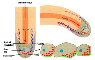
The cells in the tips of the plant rootlet respond positively to gravity, called gravitropism (the older word for it is geotropism). If you lay a growing plant on its side, the roots will respond by growing (turning) toward the gravity within 10 minutes. The mechanism for this stimulation involves tension and a plant hormone called auxin.

Auxin is a growth hormone that gets redirected
in the growing plant root. The statoliths settle
and trigger the hormone to some cells more than
others. Auxin means ”to grow” in Greek, but in
some cases, like in gravitropism of roots, it
actually inhibits growth.
The root cap (the cells at the tip of the root) have some specialized cells called statocyte (stat = position, and cyte = cell). Inside the statocytes are dense granules called statoliths (lith = stone). The statoliths are made of densely packed starch and are a specialized type of organelle called an amyloplast, which is used in many plant cells for storing carbohydrate in the form of starch (amylo = starch). The statoliths are denser than the cytoplasm of the cell; they don’t just float around, they settle out according to gravity.

Since the statoliths are connected to the membrane of the cell by the cytoskeletal actin molecules, so when they settle toward gravity, some cells in the membrane are stretched and some are compressed. This tension signals the cells to change the number of receptors for the growth control hormone auxin. More tension (more stretch) causes the auxin to move away, toward cells that are under less tension. Auxin prevents cell enlargement and cell division, so those root tip cells on the bottom receive more inhibition. Those on top enlarge more and divide more, so the root turns down. If the root is already vertical, the tension is equal in all directions, and the growth is equal in all directions – the root gets thicker and longer.

Gravitropism is related to photosynthesis in that both mechanisms involve chloroplasts, sort of. Root cells don’t perform photosynthesis, they are underground, so they don’t have chloroplasts. But they do have the amyloplastid statoliths, and these are related to chloroplasts.

Both amyloplasts and chloroplasts are specialized versions of the plant organelle called the plastid. We asked last week about what defines a plant cell – maybe the plastid is it. All plant cells have some plastids, but in different plant cells they may take different forms, including chloroplasts, chromoplasts, leucoplasts, amyloplasts, elaioplasts, or proteinoplasts, but they all start out as proplastids (pro = early and plastos = form in Greek).

Proplastids are in every new plant cell. From there
they can differentiate into other forms, including
the chloroplast. Other plastids are used for storage
or biochemical production. We will talk about statoliths
again when we discuss proprioception.
When a cell divides, each daughter gets its share of proplastids, and then depending on the chemical signals that the daughter cell receives, the proplastid will differentiate (from latin, means to make separate) into the types of plastids that the cell needs. A proplastid can become any type of plastid, and from time to time can change between forms as the plant cell requires. Think of it as a sort of stem cell inside a plant cell – if the cell happens to be in the stem of the plant, it could be a stem cell inside a stem cell!

Proplastids become etioplasts, chloroplasts or leucoplasts. The etioplast is a sort of pre-chloroplast; a chloroplast without chlorophyll. It is waiting to be stimulated by light energy before it decides to spend all the energy it requires to make the chlorophyll. The old science fair project about growing bean plants in the dark demonstrates the etioplasts. The plants are white when grown in the dark, but bring them into the light and they soon green up. The sunlight stimulates the etioplasts to make chlorophyll, become full-fledged chloroplasts and start photosynthesizing.

This is a photomicrograph of the plastids of a
red flower petal. The chromoplasts hold the
xanthocyanin pigments, but we see it as a
continuous color because they are so small.

If the proplastid does not differentiate toward a chloroplast pathway (etioplast too) then it will become a leucoplast (leuko = white). The leucoplasts don’t have color; they become specialized for the storage of plant materials. If they store starch, they are called amyloplasts. Lipid storing leucoplasts are called elaioplasts, while protein storing plastids are called proteinoplasts. Each type serves a crucial purpose in the cells they inhabit, and they can all interchange, depending on the conditions the plant cell finds itself in.

Even more important, leucoplasts that are not serving as storage organelles have biosynthetic functions. They work in the production of fatty acids and amino acids. Amino acids link together to from proteins, so their synthesis is very important for plants. Plants must manufacture every amino acid it needs, whereas we get many of ours in our diet. There are even some amino acids that humans can’t make, called the essential amino acids. Of the twenty common amino acids, nine of them must be taken in through our diet, and some people with pathologies can’t make up to seven more. Plants don’t have this luxury; all their amino acids must be made on site. Good thing they have leucoplasts.

There is one other type of plastid that we haven’t talked about, the one that is important for the Autumn tourist trade. Etioplasts and chloroplasts can differentiate into chromoplasts, organelles that store pigments (colored molecules) other than chlorophyll. Chlorophyll provides energy through photosynthesis, but they also have a cost. The old saying, “It takes money to make money” applies to plants as well. It takes energy to make chlorophyll, so it only pays to make chlorophyll when there is ample sunlight to put through photosynthesis. When the days get shorter, the profit margin for producing chlorophyll goes down, so the plant just stops making it.

Twin females were imaged after a lifetime of smoking or non-smoking.   
Can you guess who was exposed to the oxygen radicals in cigarette
smoke her whole adult life?
The oxygen produced in plant cells during photosynthesis can damage many molecules; oxygen likes to react with other compounds and steal or donate electrons. This oxidative damage can wreak havoc with the cells, just look at the face of a long time smoker – the damage and aging process from the oxidants in cigarette smoke will be evident. The chromoplast pigments, like carotenoids (oranges and yellows) and xanthocyanins (reds and purples), can serve as antioxidants, and protect the other cell structures from the damaging effects of oxygen.

So the chloroplasts lose their chlorophyll in autumn and could be called leucoplasts, but the chromoplasts still have the pigments that had been masked by the greater number of chlorophyll molecules. The trees turn magnificent colors and bring people from the cities to stay in bed and breakfasts, and to purchase handmade scarves and way too much maple syrup and apple butter. Economy and biology are so often interrelated.

Plastids are the quintescential plant organelles – no plant cell is without them in some form (well O.K., there is one exception, we’ll talk about that next week). But that still doesn’t mean that they define a plant cell. Remember that algae are not plants, but they have chloroplasts, and chloroplasts are one type of plastid. There is even a bigger exception in this area; some of the apicomplexans.

Certain protozoal organisms, including the malaria parasite (Plasmodium falciparum) contain an organelle called an apicoplast. P. facliparum or its ancestor obtained an algae cell by secondary endosymbiosis (the primary endosymbiotic event was the algae taking in a cyanobacterium), so the apicoplast has a four, not two, membrane system.

The apicoplast of the malaria parasite is of plastid
origin, but it undergoes some unplant-like changes
during cell division. Image D with the branched
apicoplast is my favorite. Those in panel F will
grow to look the one in panel A.
The apicoplast does not perform photosynthesis; we aren’t exactly sure what it does – but it is crucial for the survival of the parasite. It is located in the front of the parasite (in the direction it moves and invades cells) and is always close to the nucleus and the mitochondrion. This suggests some role(s) in energy production and molecule synthesis.

There is evidence that the apicoplast works in fatty acid and heme synthesis, like the leucoplast or in the production of ubiquinones that are important for the electron transfer chain in the mitochondria. There is also evidence that it is involved in FeS cluster production, like the hydrogenosome and mitosome. Both of these pieces of evidence show the interelationships of the endosymbiosed organelles and the connection between energy production and energy use. Whatever their functions are, if you destroy or inhibit it the malaria bug dies. As such, it has been a popular target for anti-malarial drugs.

Malaria parasites cured of their apicoplasts (cured means freed of) do not die right away. They just can’t invade any new cells and therefore can’t complete their life cycle. This is why anti-apicoplast drugs may be a boon to malaria treatment. The biosynthetic pathways in the apicoplast are the targets of four recent drugs, but the primary way to stop malaria remains the mosquito net. There is strong hope that a new vaccine, called RTS,S is a light at the end of the tunnel for this killer of millions.
The melanosome and the plastid have more in common.
The very rudimentary eye of some dinoflagellates
(dinos = rotating, and flagellum = whip) has a melanin-like
molecule in the pigment cup and the structure is called a
melanosome. However, it is of plastid orgin. The picture
above is of Polykrikos herdmanae. It has 8 transverse flagella,
as well as the pigmented eyespot to detect light sources.


One final thought on the plastid – an addition to the exception of melanosomes. We discussed a few weeks ago that melanosomes were the only organelles that could move from cell to cell. Well, that isn’t exactly so. I held off on adding the plastid to that list until we had discussed what a plastid was.

A 2012 study at Rutgers University tested whether plastids and mitochondria could move between plant cells. There results showed that entire plastid genomes could be seen in recipient cells, and the fact that the whole chromosome passed indicated that the plastid was probably moving from cell to cell intact. But there was no movement of the mitochondria, so it is a plastid (and melanosome) specific event.  The researchers hypothesize that this may be a way for plant cells to repopulate damaged cells with working organelles. As such, it would be similar to how mammalian stem cells can move mitochondria into damaged cells during tissue repair. But that is another story.

We have repeatedly talked about how the mitochondrion and plastid can replicate on their own and then are portioned out to the daughter cells when a parent divides. Can it really be that simple? I’ll bet there is a definite mechanism, and I bet that mechanism has exceptions. Let’s look into this next time.

Gregory Thyssena,Zora Svaba, and Pal Maligaa (2012). Cell-to-cell movement of plastids in plants Proc Natl Acad Sci U S A. , 109 (7) DOI: 10.1073/pnas.1114297109

For more information or classroom activties on plastids, gravitropism, or Plasmodium falciparum see:

Plastids –

Gravitropism –
207.62.235.67/case/biol215/docs/roots_gravity.pdf

Plasmodium falciparum

Thursday, November 9, 2017

The Evolution Of Cooperation

Biology concepts – biological timeline, serial endosymbiosis, endocystosis, evolution


Taxonomy, the placing of species in different
groups based on their characteristics, changes
everyday – literally everyday – organisms are
placed in different groups and groups are created
and eliminated. That better be a temporary tattoo!
If we look at the 3.5 billion year history of life on Earth, we see that out planet was lifeless for almost a quarter of its span, and animals have been around just a short blip of time, a mere 760 million years. Often, it seems that the big numbers to get in the way of understanding the time line as a whole.

If we treat the entire history of earth as one year, we might get a clearer picture. Earth coalesces from space dust on January 1st, but it isn’t until March 22nd that we find the first evidence of life. These most primitive fossils are of the prokaryotes called Archaea (Greek for “ancient”). Not long after this, maybe a week or so, the eubacteria and Archaea separate from one another.

Then we have to wait until August 7th to find a big change; the first eukaryotic organisms are seen. These represent a fundamental change in the organisms, having nuclei and membrane bound organelles. It's amazing that we must travel 3/4 through our one year time line before we see a cell that looks somewhat like ours!


Here is one of the Namibia sponge fossils recently
discovered in Africa. It represents the oldest animal
in the fossil record. Just how that was recognized as a
fossil is beyond me – I think I have six of those in my
garden!
Later in the year, around October 30th at noon, we see the first animals. Fossils of Namibia sponges in Africa were first reported in February of 2012. This fossils are 100 million years older than the previously oldest animal remains, so our new data means that animals have been around for an additional week in our time line of a year.

Insects appear about Nov. 26th, while mammals first show up around Dec. 8th. The dinosaurs became extinct sometime in the afternoon of Dec. 26th, so they had very little time to play with their Christmas presents. Homo sapiens (us) didn’t appear on the doorstep looking for holiday cheer until 11:40 pm on New Years Eve, Dec. 31st!

Our time line analogy shows us that prokaryotes are the wise old ancestors; we aren’t even old enough to be rebellious teenagers, although we still think we know everything. The key question is: how did we progress to analogy-makers from single celled Archaea? If we put together several of the topics we have been discussing in the past three weeks, we may come up with an interesting step in the process. Our clues include:

1) Microcompartments exist in bacteria, like organelles, and they also exist in eukaryotic cells, especially in nucleus' function. This links eukaryotes to prokaryotes.

2) Sometimes cells will engulf objects, parts of other cells, or other cells. Depending on the size of the particle or cell, we may call this endocytosis or phagocytosis, and is similar to how we saw keratinocytes take up melanosomes.

3) Three eukaryotic organelles, the nucleus, the mitochondria, and the chloroplast have double membranes, and they each have their own DNA.

4) There are two different types of prokaryotes, archaea and bacteria.

Bacterial microcompartments give prokaryotes some compartmentalization in order to carry out necessary chemical reactions. Eukaryotes also have some prokaryotic microcompartment remnants, like the nuclear vault complex. This shows crossover between prokaryotes and eukaryotes, and gives us clues about eukaryotic origins. In fact, the currently accepted theory about the evolution of organelles - the very thing that makes cells eukaryotic - has to do with both types of prokaryotes - archaea and bacteria.


There are three types of endocytosis (with exceptions).
Endocystosis of large objects and cells is called phagocytosis.
Internalization of very small molecules and fluid is called
pinocytosis. Other molecules of various sizes have specific
receptors that recognize them on the cell surface. They are
brought in by receptor-mediated endocytosis. Notice that no
matter what method is used, the internalized particle ends up
surrounded by part of the cell membrane.
The key to their interrelationship has to do with endocytosis (endo = into, cyto = cell). Most prokaryotic and eukaryotic cells eat other cells; they do it all the time – it is how heterotrophic organisms (those that can't make their own carbohydrates, ie. non-plants) gain their nutrients. We do it too, just on a larger scale; we eat millions of cells at a time; often these millions of cells can take the shape of a steak or a carrot.

When a cell, protein, other molecule is engulfed by another cell, it is wrapped in a portion of the aggressor cell’s membrane. The naked molecule is now contained in a vesicle, a membrane bound sac, like the melanosome. If the endocytosed material is an entire cell, something that has its own membrane, then it ends up with two membranes, just like the mitochondrion, chloroplast, and nucleus.

Most often, when one prokaryote phagocytoses another, the story is over….gulp, yum, digest. But scientists believe that long ago (sometime in the first week of August in our time line) an endocytosed cell did not go gentle into that good night. Instead, it took up residence in the cell that ate it. In this rare case, it turned out that both cells gained from the situation.

The endocytosed cell was protected from other predators and had a ready supply of nutrients from the parent cell. The captured cell made lots of ATP, but it didn’t need much because it was being supplied with everything it needed; it didn't need to make energy to move or hunt or escape. Most of its ATP production went unused. Perhaps it moved this excess ATP out into the parent cell. So the parent cell gained a source of ATP production. This was mutualism, a type of symbiosis in which both parties benefit.


Clownfish clean the sea anemone and keep it
parasite free. The poisonous anemone provides
a safe environment for the clown fish; no
unwanted house guests! This is a good example of
mutualistic symbiosis. Bet you didn’t know you
learned things from Finding Nemo.
Imagine if the same thing happened with a cyanobacterium, a cell that could perform photosynthesis. The same sort of symbiosis might be set up, with the endocystosed cell providing carbohydrates and the parent cell providing protection.

Now imagine that these captured cells, the photosynthesizer and the ATP maker, replicated themselves inside their parent cells just as they would if they were outside, living on their own. They could easily do this since they still retained their own DNA and cell division mechanisms.

This is in fact what scientists believe happened. The endocytosed cells that produced extra ATP evolved into our mitochondria. Endocytosed cells that could do photosynthesis became the chloroplasts of plants. Not all cells are plants because not all cells with an ancestral mitochondria also ate a cyanobacterium. The fact that plants cells have mitochondria as well as chloroplasts tells us that plant cells developed AFTER cells with mitochondrial ancestors.

But the nucleus may be a tougher nut to crack. It may be that an endocytosed cell good at keeping DNA safe and producing ribosomes became the nucleus, by endocytosis. The data suggests that our DNA is closer to archaeal DNA than bacterial DNA, so it would have been a eubacteria endocytosing an archaea. Or perhaps the archaea invaded the bacterium rather than being endocytosed. The nucleus does have a double membrane and uses some prokaryotic microcompartments to this day, so this could make sense.

But other theories also exist, including one that says an intermediate eukaryotic cell, theoretically called a chronocyte, had developed some organelles on its own or by endocytosis, including a cytoskeleton. This internal structure allowed the cell become bigger, and engulf a cell large enough to evolve into the nucleus.

Another theory uses an evolutionary exception as its basis. Some aquatic bacteria, called planctomycetes (planktos = drifting and mycete = fungus-like), have an organized interior, with something that looks like a nucleus with pores, called a nucleoid. In fact, when they were first discovered, planctomycetes were mistaken for small fungal cells. However, we know they are prokaryotes by DNA sequencing. I thought prokaryotes didn’t have nuclei! Remember that in biology, there is almost always an exception. The planctomycete nucleoid structure suggests that the nucleus may have evolved on its own, without endocytosis.


The planctomycete species, Pirellula (latin for small pear),
is an exceptional bacterium. It has a primitive nucleus
and a stalk that makes it look like a eukaryotic
fungal cell. It was misidentified for a long time, and is
a prime example of why the tattoo above was a bad
idea!
Finally, another theory posits that the nucleus originated from a virus infecting a primitive prokaryote, and this internalized virus forming a nucleus or causing the cell to be predated by another cell. Even though there are different theories for the nucleus, we can see that the three organelles that have double membranes look like they could have been endocytosed cells, that then evolved into the organelles we see today. Endocytosis resulted in symbiosis, so the theory of organelle development is called endosymbiosis.

Endosymbiosis is a cool idea and has lots of support. Besides the double membrane evidence, lets look at how dividing cells get more mitochondria and chloroplasts. These organelles replicate on their own by binary fission, just like bacteria. They can replicate on their own because they have their own DNA. Mitochondrial DNA (mtDNA) and chloroplast DNA (chDNA) are smaller pieces of DNA than nuclear chromosomes, mtDNA and chDNA look much like the small genomes of bacteria. They are also circular pieces of DNA, not linear like our nuclear chromosomes.

By replicating through binary fission, they can be portioned in the dividing cell so that each daughter gets some of these crucial organelles. But it isn’t as if mitochondria and chloroplasts of today look just like the engulfed ancestors. Mitochondrial and chloroplast genomes are greatly reduced from what they used to be.


Serial endocytosis is also called secondary (2˚) endocytosis.
This refers to the movement of DNA from internalized
cells to the nucleus of the endocytosing cell by lateral
gene transfer. This strengthens the symbiotic relationship
between the two organisms until they can be considered
one total organism.
The mitochondria only codes for about thirteen proteins, just enough for it to replicate on its own. The DNA that codes for the rest of the 1500 or so proteins needed for mitochondrial function have been transferred to the nucleus over time. For a discussion of the chloroplast and its horizontal gene transfer to the nucleus, see the posts on C. litorea, the photosynthetic sea slug.

We know that these gene transfers were actual events based on the structure and nucleotide ordering of the mitochondrial and photosynthetic sequences in the eukaryotic chromosomes; they are structured and coded in ways that are typically bacterial. Because of this slow transfer of DNA to the nucleus, endosymbiosis has evolved over time, changing again and again until we got today’s organelles. Therefore, our idea of organelle development is sometimes called serial endosymbiosis theory (SET), because it must have had several different changes through evolution.

Now that we have laid out the evidence and sense for the serial endosymbiosis theory, next week we can talk about some exceptions that show us that that some organisms just can't stick with something that seems to work. Some life just has to take the road less traveled.


Okie JG, Smith VH, & Martin-Cereceda M (2016). Major evolutionary transitions of life, metabolic scaling and the number and size of mitochondria and chloroplasts. Proceedings. Biological sciences / The Royal Society, 283 (1831) PMID: 27194700

Kostygov AY, Dobáková E, Grybchuk-Ieremenko A, Váhala D, Maslov DA, Votýpka J, Lukeš J, & Yurchenko V (2016). Novel Trypanosomatid-Bacterium Association: Evolution of Endosymbiosis in Action. mBio, 7 (2) PMID: 26980834

Erbilgin O, McDonald KL, & Kerfeld CA (2014). Characterization of a planctomycetal organelle: a novel bacterial microcompartment for the aerobic degradation of plant saccharides. Applied and environmental microbiology, 80 (7), 2193-205 PMID: 24487526


For more information or classroom activities on history of life time lines, endocytosis,  serial endosymbiosis theory, evolution of eukaryotes, or planctomycetes, see:

History of life on Earth timelines -

Endocytosis –

Serial endosymbiosis theory –

Evolution of eukaryotes –

Planctomycetes –