Showing posts with label protein. Show all posts

Wednesday, January 27, 2016

An Infectious, Genetic Disease? Better Sleep On It.

Biology concepts – thermoregulation, sleep, genetic disease, infectious disease, central dogma of molecular biology, form follows function


Even rats have to get some sleep. It was nice to have the sleeping cap,
but unnecessary for a sleep deprivation study. Not a good use of
research dollars.
“I’m dying for a good night’s sleep.” Is this just hyperbole, or an impending warning of death? For laboratory rats, sleep deprivation does kill. During their insomniac downward spiral, the rats tend to get hot and can’t cool down – you know, they can't thermoregulate (see Can’t We Just Go With The Flow). This doesn’t mean that a loss of the ability to thermoregulate is what kills the rats, but it does suggest a connection between sleep deprivation and the hypothalamus.

We looked at the hypothalamus in our story of endothermy. This evolutionarily old brain structure implements a set point temperature for the body and receives information about the temperature of different parts of the body. When the body temperature deviates from the set point, the hypothalamus initiates bodily mechanisms to normalize the temperature.


Apparently one of the effects of sleep deprivation is that you
become semi-transparent.
People with severe insomnia tend to sweat more and have higher core temperatures even though they say they are cold. They also have extreme high blood pressure, pulse, and appetite. These symptoms suggest that sleep deprivation messes with the hypothalamus, since functions of the hypothalamus include themoregulation, sleep, hunger, thirst, reproductive readiness in females, and stress responses. What scientists don’t know yet is just how sleep deprivation actually kills the rats or harms people.

Dying from a lack of sleep is not just a rat problem, a few very unlucky humans die from it as well. Fatal familial insomnia (FFI) is a very rare genetic disorder; it has been reported in only 40 families worldwide. Before describing the truly horrible way these patients die, let’s look at what causes the disease.

FFI is caused by a point mutation in the gene for the prion protein PrPc. A point mutation means that one nucleotide on the DNA is changed, which leads to a change in the protein coded for by the DNA. Three unit (nucleotides) segments of the RNA (made from the DNA template) work together (called a codon) to code for one protein building block (amino acid). In the case of FFI, the amino acid called aspartic acid is changed to one called asparagine, and this changes the protein’s shape. 


The left image shows mRNA bases recognized in sets of three
(codons) by tRNAs with amino acids attached (Ser = serine, tyr =
tyrosine). The amino acids are linked to because proteins. The
lower section is the genetic code, showing which amino acids are
coded for by which codons. The right image shows how proteins
fold. The primary structure is the amino acid sequence. The
secondary structure comes from interactions of adjacent amino acids,
including spirals called helices or sheets. The tertiary structure comes
from the folding up of the entire protein, while the quaternary
structure comes from the interaction of different proteins into a
larger complex.
PrPc is made up of 250 amino acids linked together in a chain. Each different amino acid carries a different shape and charge and will interact with every other amino acid differently. The sequence of amino acids in a protein cause it to fold into a specific shape. It is the protein’s conformation (shape) that determines its function. This is the opposite of what we determined for evolved organism characteristics, where form follows function (see Do You Have To Be Ugly To Hear Well?). With proteins – function follows form!

Mutation of that single amino acid at position 178 (aspartic acid is negatively charged, while asparagine is positive) causes the folding, and therefore the function, of the protein to change. Aspartic acid is sometimes abbreviated "D", while asparagine is called "N"; therefore, the mutation is often indicated as D178N (D at position 178 is changed to N).

Many genetic mutations result in no change in amino acid, or a change that bring a large enough change the shape to cause a change in function. But when it does, good or bad things can happen. On one hand, the altered protein might confer an advantage to the organism, one that promotes survival in the environment or after an environmental change.This positive selection through reproductive advantage become the new normal – and this is evolution

On the other hand, the change in amino acid sequence, form, and function could be destructive. Disease might be the result, or perhaps a change in the organism that reduces reproductive success. One of these two results is what occurs with the FFI mutation of the prion protein.

When the mutated prion folds differently, it forgets its day job and moonlights as a sinister evil force. Every other prion protein it contacts, WHETHER MUTATED OR NOT, is coaxed into changing its shape. The new prions turn to the dark side, then change other prion proteins they contact, multiplying the effect. The poorly folded prion proteins will stick together, come out of solution, and form solids (plaques) where they settle out. In different prion protein diseases, this settling out occurs in different parts of the brain. In FFI, it is the hypothalamus.


In the top image, the PrPc on the left is properly folded. The green
represents alpha helices and the blue arrows represent beta-pleated
sheets. The right image shows the malfolded version of PrPsc. It is a
tighter structure, which partially explains why protein-degrading
enzymes don’t work on it. . The lower cartoon shows that the PrPsc
can force the PrPc to assume the improper form, and these then
aggregate into plaques.
The prion plaques are longer lived then the regular prion protein; normal cellular enzymes whose job it is to degrade proteins won’t work on prion plaques. And worse, if some of the malfolded protein is transferred to another animal, the recipient will develop plaques and disease as well. That makes this an infectious disease that isn’t caused by a bacteria, fungus, parasite, or virus. The prion is an infectious protein! What a terrible exception to the rules of infectious diseases.

We see here a protein that can replicate itself (not by building more of themselves, but by changing the form of normal proteins), and that makes it a repository of biologic information. This is an exception to the central dogma of molecular biology, which says that DNA is the sole information storing material.

FFI moves from person to person through heredity, but if a non-affected person comes into contact with some brain material from an FFI patient and that material entered their bloodstream, it can be transmitted this way as well. A prion protein disease called Kuru is famous for being transmitted from person to person.

The Fore tribe in Papua New Guinea once observed a ritual wherein they honored a dead tribe member by eating part of their brain (called ritualistic mortuary cannibalism - gasp!). Because of this, there was an epidemic of Kuru in this tribe in the early 1900’s. Over a period of 3-6 months victims would become unsteady, irrational with bouts of laughter, and then degrade mentally and physically to the point of death. There are more than twenty known prion diseases (mad cow disease, Creutzfeldt-Jakob, scrapie, etc.), and Kuru suggests that some might have no genetic component, only person to person transmission.


A member of the Fore tribe is shown on the left. This tribe used
to celebrate the lives of departed members by eating their brains.
This spread a prion protein disease called Kuru, a protein disease
that is infectious! The Fore tribe still lives in Papua New Guinea,
although there are fewer of them than before Kuru.
The differences between the various prion diseases are based on the specific prion protein mutation, what part of the brain is attacked, and how potent the prion is at refolding normal prion proteins. For instance, the D178N mutation in FFI also occurs in Creutzfeldt-Jakob Disease (CJD), but a normal polymorphism (an amino acid change that doesn’t change form or function) at position 129 determines the fate. If amino acid 129 is methionine, the the person gets FFI, if it is valine, then they get CJD. 

The families that suffer from FFI have the D178N mutation, and also pass on the polymorphism for methionine (M) at position 129. Even more gruesome, some cases of prion protein diseases can be sporadic, not associated with either an inherited mutation or transmission. The malfolded prion can very rarely arise out of nowhere in isolated individuals.

The mutated PrPc is passed on via inheritance. You get one copy of each chromosome from each of your parents, so for an individual gene, you might get two normal copies, 1 mutant copy and 1 normal copy, or 2 mutant copies. Some diseases require that you must inherit two mutant copies for symptoms to show (recessive), but other require only one mutant copy (dominant, it dominates the trait from the other parent).

FFI is autosomal dominant (not associated with the X or Y sex chromosomes), so the chance of getting a mutant copy and the disease if one parent has it is 1 in 2; these are bad odds. But, if everyone with FFI dies, then why is the disease still showing up in families. Remember that we said above that some genetic diseases can, but don't have to, affect reproductive success. Unfortunately for those with FFI, the symptoms appear in the victims’ fifties, after they have had children. Natural selection doesn’t eliminate FFI from the population because FFI doesn’t appear affect reproduction.

The first symptoms of FFI include sweating while feeling cold. Later, the ability to get a good night’s sleep is lost, followed closely by the inability to nap. As the disease progresses, there are panic attacks, phobias, and no sleep whatsoever. After 4-6 months, mental abilities start to degrade. In its final stages unresponsiveness precedes death. 

This is especially sad way to die, because during the majority of the disease course the patient is aware of everything going on. At least with middle to late Alzheimer’s disease the patient is blissfully unaware of their dementia.


For both the gross and microscopic images, the left example is from prion protein disease victim, while the right example is from a normal brain. The brains on the left show how great the loss of tissue can be in Creutzfeldt-Jakob disease. The microscopic image from the diseased brain shows the plaques and the resulting holes in the brain structure. The small gaps in the normal brain on the right are a result of shrinking of tissue after it was on the slide.
On autopsy, the hypothalmus of an FFI sufferer looks like it has been hit with a shotgun blast. Holes are present in the tissue, representing areas where neurons have been lost due to inflammation and triggered cell death. The affected area of the brain takes on a spongy appearance, so prion protein diseases are lumped together and called transmissable spongiform encephalopathies (encephalon = brain and pathy = disease). Unfortunately, there are no cure, treatments, or vaccines for any of these prion diseases.

It is the hypothalamus' control of sleep cycles and circadian rhythms that promotes survival in animals. But what about plants? They don’t have a hypothalamus. Can they suffer from loss of circadian activity? In a word – yes!  And this will be our starting point next time.


For more information or classroom activities on prion proteins, central dogma, infectious or genetic disease, the genetic code or protein structure, see:

Prion protein and diseases –

central dogma of molecular biology –

infectious disease –

genetic disease –

genetic code –

protein structure –
nwabr.org/sites/default/files/learn/bioinformatics/AdvL5.pdf
 

Wednesday, July 2, 2014

How Do Mosquitoes Find You?

Biology concepts – semiochemicals, hematophagy, proboscis, thermosensing, TRPA1


Sure, mosquitoes suck blood and pass along malaria
that kill more humans than any other infectious disease.
But would it be good to get rid of them. They provide
food for birds – one scientist suggests that elimination
of Arctic mosquitoes could reduce northern bird
populations by 50%. And mosquitoes pollinate flowers
too, like blueberries and cranberries. See, they’re
not all bad.
We can start our summer series of biology questions by continuing our discussion of taste and thermosensing. It seems that some people are bitten by mosquitoes if they peak out the front door, while other people can sit outside next to tall grass or ponds for hours with suffering a single bite. Unfortunately, I happen to be in the former category.

How do mosquitoes find some people and not others? Are some people just tastier than others?

First let’s get some common misconceptions and basic information out of the way. Do mosquitoes bite you (or any other animal)? No, they have no teeth, so they don’t bite in the traditional sense. What they do have is an elongated set of mouthparts called a proboscis. The sheath on the outside retracts as the longer parts inside pierce the skin like a hypodermic needle. Only this is a flexible hypodermic needle, small enough to go around individual cells and look for a small vein or venule.


On the left is a drawing of the mosquito proboscis parts. Most sit
in a groove of the labium, which retracts as the rest is injected
into the skin. The maxillae and mandibles are like our upper and
lower jaws. They are the sharp parts. The hypopharynx is what
delivers the anticoagulant saliva. On the right is the parts put
together. The fuzzy part is the labium and the sharp tips are
from the maxillae.
Take a look at these videos taken from a 2012 PLoS One study of a mosquito biting a mouse. The squarish objects are skin cells, and the red streaks are blood vessels. The second video in the sequence shows what happens when the proboscis finds a vessel and starts to suck out the blood. Makes you respect the mosquito a bit more – these are some determined females.

Of course it’s only the females that feed on blood. This suggests that feeding on blood is related to having babies. And it is – just not in a “gotta get the baby some food” sort of way. Most mosquito species require a blood meal in order to develop viable eggs. Females get energy from drinking nectar (full of carbohydrates), but they need protein to produce yolk for the eggs. They get the protein from feeding on blood. If the female doesn’t feed on blood, the eggs will be produced, but they won’t be able to hatch and become larva.

But here is one of our exceptions – some mosquitoes have gotten around the need for blood meals. All 92 species of mosquito in the genus Toxorhynchites (elephant mosquito) don’t need to feed on blood. Instead, their larvae feed on the larva of other mosquitoes, and the gather the proteins they need to lay viable eggs from their larval meals. They store the amino acids in their larval and pupal bodies, until they become adults and need them to lay eggs of their own.

Compare the sizes of the elephant mosquito (left) and
A. aegypti.  I’m very glad that the females of the
Toxorhynchites genus don’t suck blood. They could drain
people dry! Even though their size is small, species like the
one on the right can consume 300 ml a day from every
caribou in a herd when they are swarming.


This suggests that the elephant mosquitoes could be used to combat disease spreading mosquitoes, like the Aedes aegypti mosquitoes that spread dengue fever, yellow fever and the current disease of interest, chikungunya fever. And the elephant mosquito has been used as a natural biocontrol agent. What's weird is that A. aegypti females actually help the situation.


A. aegypti, and many other mosquitoes that lay eggs in water, have larvae that eat bacteria. So they want to lay eggs where there are a lot of bacteria. Well, the eating of larvae by Toxorhynchites species leaves lots of little pieces of mosquito larva in the water, and this provides bacteria with a lot of food. A June 2014 study showed that A. aegypti females actually prefer to lay eggs in water that contain predators for their larva, because it increases the bacterial numbers so much. Thos that survive have lots of bacteria to feed on. It’s a calculated behavior – risk being eaten or risk starving.


So some mosquitoes will go a long way and risk death in order to get a good meal for their potential offspring. They’re looking for mammals usually, but even here there are exceptions. Some mosquito species, like Culiseta melanura, feed almost exclusively on bird blood – they say it tastes like chicken.


The picture represents the transmission cycle for
eastern equine encephalitis virus (EEEV). It can’t be
transmitted from mammals to other animals, so they are
called dead-end hosts. But it can produce disease in
them. Humans most often will present with a limiting or
subclinical disease, but horses have a hard time with it.
The major source is in birds, and the transmission from
bird to bird is by mosquitoes that rarely bite humans. The
way into dead-end hosts is by a mosquito that normally
bites mammals occasionally biting a bird, or the rare
occasion that a bird specific mosquito will bit a mammal.
But just because they feed mostly from birds doesn’t mean they aren’t important disease transmission. They are – for horses. Eastern equine encephalitis virus is passed from bird to bird by C. melanura, so the birds, especially cardinals, are a reservoir of virus. Then, when another species of mosquito that is less particular about its host species bite a bird then bites a horse or person, the disease can be spread. There are even cases where a C. melanura will occasionally feed on a human and spread the disease directly.

With this background, we still need to answer our question of the day – how do mosquitoes find a blood meal. Believe it or not, your socks help answer the question. For many years it was assumed that mosquitoes followed the heat of warm-blooded animals in order to find a meal, but this was an assumption that was not tested rigorously.

Then it was discovered that carbon dioxide (CO2) is a strong cue for mosquitoes seeking sustenance. CO2 means respiration, and respiration possibly means mammals. The mosquitoes have taste receptors in their antennae and mouths that will sense changes in CO2 and they will follow the path of more carbon dioxide right to your nose and mouth (see this post).

Large people and pregnant women tend to exhale more CO2, so they will be more attractive to mosquitoes. But there are large individuals who never get bothered by mosquitoes. Maybe there’s more to it.

Semiochemicals are part of the answer. Semio- comes from the Greek meaning signal, like in semaphore flags. So semiochemicals are molecules emitted by organisms for communication. Pheromones are the most famous of the semiochemicals – and we know that these are used in many animals, from helping to guide ants to follow the path of their predecessors, to influencing mate choices in many animals.

Semiochemicals might be attractants or repellants. In some cases, they can be both. Take human body odor – it contains dozens of semiochemicals, people find body odor repulsive, but mosquitoes enjoy it like the smell of fresh apple pie. Of course, body odor is only offensive nowadays; before the advent of deodorant, daily or three times daily baths and showers, perfume, aftershave, and of course Axe products – everybody smelled like that guy that lives under the bridge.


On the top of this image is a general idea of semiochemicals.
If they work on members of the same species (like mating
signals), then they are called pheromones. If they work on
other species, they are called alleochemicals. Each can be
either attractive or repellant. On the bottom is a homemade
mosquito trap. You might be able to see that it has been
baited with old shoes and grimy socks.
Bacteria feed on the sweat, sugars and proteins that mammals exude, and they give even more semiochemicals. This can make you more or less attractive to mosquitoes. In general, people with many types of bacteria on their skin are less attractive, while those with mostly a few attractive species will get bitten more often. Having a high number of bacteria is a turn off too, probably because that would expose the mosquitoes to more possible pathogens as well. Is it possible to be so disgustingly colonized that even mosquitoes won’t land on you?

Mosquitoes are attracted to several different semiochemicals, including octenol, CO2, and nonanal. On mosquito antennae, especially the female antennae, there are receptors in the sensilla (see this post) for at least 27 different chemicals in human sweat.

Studies have shown that old socks are a good experimental attractant for mosquitoes. Instead of using an arm or other body part, scientists will compare the attractive ability of someone’s old sweat socks to individual chemicals or mixtures of chemicals. Of course, whose socks you use matters as well. Some people are classified as high attractors (HA) and some as low attractors (LA), so studies often include comparisons of chemicals or mixtures to both HA and LA socks.

But there are other considerations as well. People with blood type O secrete different semiochemicals and are more attractive to many species of mosquitoes. Go ahead, try to change your blood type so you’re less attractive to mosquitoes.

Different species may aim for different body parts. Some seem to prefer feet and ankles, but this may be because they are closer to the ground. If convection currents created by the body heat rising suck the mosquitoes in from below, then it is really the fact that they are following their noses and not going after feet particularly. A small 1998 study showed that mosquitoes that went after feet and ankles preferentially did not do so when the volunteers lied on their backs and raised their feet high in to the air. But, what we have stumbled across in this discussion is body heat.


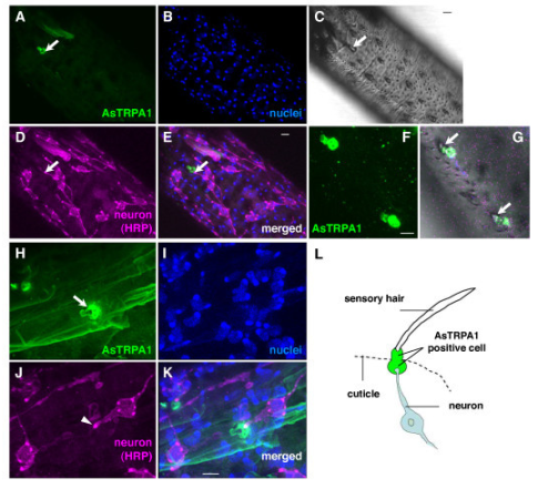
This is part of a complex figure from a 2011 scientific paper.
In addition to the pretty colors used, the message is that these
researchers identified TRPA1 ion channels on the proboscis
of a species of mosquito. They don’t just sense heat with
their antennae, but also their sucking parts. I wonder if the
interior parts also have TRPA1 to help them find a vessel
when the proboscis is inserted into the tissue.
But what was old is new again…. Scientists are again looking at heat as an attractant for mosquitoes. As compared to HA or LA socks, heat isn’t a strong attractor, but warm socks attract more mosquitoes than cold socks. On the other hand, a 2010 study says that heat and moisture is a greater attractor than heat alone, so it would seem that people working outside in the heat would be the perfect attractors for mosquitoes.

Since heat does seem to be an attractor, it would follow that female mosquitoes would have a receptor for heat. Voila, a new study shows that mosquitoes have sensilla on their antennae and palps that house TRPA1 ion channels. A 2011 study even showed that one malaria-carrying mosquito has TRPA1 receptors on its proboscis. We have talked before about how many mammals use this receptor to sense noxious cold as well as chemicals that cause irritation or pain.


On the left is a species of tick. You wouldn’t believe how big they
can get when feeding on blood. Look it up – I dare you. On the
right is a bedbug. The bedbug is not that closely related to the
tick, since the tick is an arachnid. Count the legs on each – spiders
(arachnids) have eight legs, insects have six. Both these animals
feed on blood, but no one has identified a heat sensor in them.
But in birds, reptiles and insects, TRPA1 is a heat sensor. The 2009 study showed that the TRPA1 were expressed on the female antennae only. But that isn’t to say that only female mosquitoes have TRPA1. A 2013 study indicates that A. gambiae mosquito larvae have functioning TRPA1 so that they can sense water temperature and stay in the most comfortable water.

So mosquitoes (most female mosquitoes) are finding suitable hosts for blood meals by using their senses of taste, smell, sight, and infrared detection. There are other vampire insects as well, ticks, bedbugs, etc. I wonder if they are using heat sensing too. These have yet to be reported on.

Next week, a related question – just how and why do mosquito repellants work?


Maekawa E, Aonuma H, Nelson B, Yoshimura A, Tokunaga F, Fukumoto S, & Kanuka H (2011). The role of proboscis of the malaria vector mosquito Anopheles stephensi in host-seeking behavior. Parasites & vectors, 4 PMID: 21272298

Albeny-Simões D, Murrell EG, Elliot SL, Andrade MR, Lima E, Juliano SA, & Vilela EF (2014). Attracted to the enemy: Aedes aegypti prefers oviposition sites with predator-killed conspecifics. Oecologia, 175 (2), 481-92 PMID: 24590205

Olanga EA, Okal MN, Mbadi PA, Kokwaro ED, & Mukabana WR (2010). Attraction of Anopheles gambiae to odour baits augmented with heat and moisture. Malaria journal, 9 PMID: 20051143

Liu C, & Zwiebel LJ (2013). Molecular characterization of larval peripheral thermosensory responses of the malaria vector mosquito Anopheles gambiae. PloS one, 8 (8) PMID: 23940815

Wednesday, November 6, 2013

Rewriting the Genetic Code

Biology concepts – DNA, RNA, tRNA, nonstandard nucleotides, codon, anticodon, genetic code, selenocysteine, isodecoder, mitochondria


Just looking the Imperial Hotel in Tokyo doesn’t really give us an idea
of why they inspired Frank Lloyd Wright’s son to invent Lincoln Logs.
 It was the interlocking beams of the basement in which his vision was
born. They were supposed to protect the hotel from earthquake
damage. It worked. In 1923, the same year the hotel was finished,
there was a great earthquake in Tokyo and the Imperial was one of
the few buildings that survived. It also survived the bombings of
WWII. So they tore it down in 1968.
In 1916 John Lloyd Wright invented Lincoln Logs. The construction set was based on his memory of the Imperial Hotel in Tokyo, an edifice designed by his father, Frank Lloyd Wright. The construction set had specific pieces that fit together in a specific way.

The first edition of Lincoln Logs, sold in 1918, gave instructions for building Abraham Lincoln’s boyhood home and Uncle Tom’s cabin. The parts were commensurate for building those structures. Each set of instructions called for the small pieces to be put together in a certain order so that the resulting product conferred a meaning – this is where Lincoln grew up or this is where Tom lived.

DNA and RNA are similarly constructed. There are a few pieces (nucleotides A, C, G, T, and U) that can be used to build different structures. Each small piece can be joined with other small pieces to become part of the whole structure, a structure with meaning. In the case of DNA and mRNA, three nucleotides in a row can confer meaning for one protein building block. The entire series of nucleotides then has the meaning of an entire protein.

The three nucleotide codons relate to a certain amino acid building block to be inserted into a growing protein. This code, the genetic code, gives meaning to the string of DNA nucleotides in genes and the string of nucleotides in the mRNA transcribed from the gene. This is usually where our learning about nucleic acids ends.


The top left picture is Marshall Nirenberg, the initial decoder of the
genetic code. The right photo is Robert Holley, discoverer of tRNA.
Below is the genetic code in graphic style. The four large letters
represent the possible first bases of a codon (in mRNA). The light
yellow letters are the possible 2nd bases, and the darker yellow letters
are the final possibilities. Outside are the amino acids that are coded
by the individual codons. Note that most have more than one codon
and some have codons that begins with different letters, like serine
at 1:00 and 8:30.
The history of the genetic code is worth knowing, as is the history of about every part of science. I often use history to illustrate points in the blog. It is said that those who ignore history are doomed to repeat it, and science has its own version of this axiom, “Six months in the lab can save you a whole afternoon in the library.” Think about it. And besides saving you from repeating others' work, knowing history helps you ask better questions.

But I digress – let’s talk briefly how we decoded the pathway of gene to protein. It begins with Watson and Crick publishing the structure of DNA in 1953. We knew how the different bases could be ordered, but we still didn’t know how they called for a specific amino acid sequence.

In 1955, Francis Crick thought he had an idea about how it might occur, but he didn’t have all the players. He called his idea the Adapter Hypothesis. What he was missing was the adapter, the piece that he said carried amino acids and put them in the correct order.

One neat trick came from George Gamow, a nuclear physicist best known for his role in theorizing the Big Bang (the birth of elements from a cosmic explosion, not the TV show). We had four nucleotides to encode information and 20 (you and I know there are 22) amino acids to be coded for. He used some “way beyond me” math to determine that the most efficient mechanism would have three nucleotides code for one amino acid.

This was followed by an interesting experiment done by Marshall Nirenberg at the National Institutes of Health near Washington, DC. He made a synthetic RNA of a single nucleotide (UUUUU….). He then combined this with the innards of a bunch of cells (cell lysate) so that everything needed to make a protein would be present. He detected a peptide of phenylalanine amino acids. What is more, there were 1/3 as many amino acids as there were nucleotides!

So UUU coded for phenylalanine. This was followed by many more experiments using different sequences of nucleotides, and the code was decoded. Along with this knowledge came the discovery of tRNA by Robert Holley in 1965. This RNA combined an anticodon sequence to recognize a codon on mRNA and carried the appropriate amino acid at the other end. The tRNA was Crick’s adapter, and perhaps the code would have discovered years earlier if the adapter had been pursued in earnest.


The process of turning an mRNA into a protein involves the ribosome and
the tRNAs. When an mRNA is bound to a ribosome, the three nucleotides
in the codon (pink letters) match with three letters of a tRNA anticodon
(blue letters). Different tRNAs will drift in and out until the right one is
bound. The tRNA has the amino acid (aa) bound to the end opposite the
anticodon. If this is the first position of the peptide, it will occur in the P
(peptidyl) site. The second tRNA will be added to the A (acceptor) site and
the ribosome will shift as it creates a peptide bond between the two aa’s.
The shift puts the first aa in the E (exit) site to reelase the tRNA, the 2nd aa
goes to the P site, and the A site is open for the next tRNA.
There are 64 possible codons that can be made from four nucleotides (4 x 4 x 4), but Holley found fewer than 64 tRNAs, one for each codon. Even I know that this kind of math doesn’t work. It turned out that the genetic code was degenerate; more than one codon calls for a particular amino acid. Most amino acids have 2-4 codons assigned to them (we have talked about the exceptions to that rule).

In most cases, codons that call for the same amino acid have the same first two nucleotides; it’s the third position (wobble position) that varies. It was discovered that the third position of the anticodon binds to the DNA very loosely, so the codon/anticodon binding is usually determined by the first two nucleotides. This allows a single tRNA to recognize more than one codon.

It turns out that there are 40-55 different tRNAs, depending on the organism. Why so many? As an example, arginine is coded for by several codons (CGG, CGA, CGC, CGU, AAC, and AAU). It is impossible for one tRNA to recognize both AAC and CGG, so there must be more than one tRNA for arginine.

Serine and leucine are like this as well, and there are most certainly some amino acids whose tRNAs can’t bind to all four possible nucleotides in the wobble position (like glycine), so they would need more than one tRNA. These are the isodecoder tRNAs (different anticodons, but code for same amino acid).

There are also different isodecoder tRNA genes, having different sequences outside the anticodon, but code for the same amino acid. Humans have about 274 genes for our 55 different tRNAs. This implies that the different sequences might have some functions other than just helping to add the right amino acid to a growing peptide sequence.

A 2010 minireview talked about those possible tRNA functions. In one discussed study, a cleaved tRNA is shown to have increased expression when cells are proliferating. Reducing the levels of this cleavage product reduced the rate of cell division. In another study, a tRNA cleavage product silenced the expression of a specific gene. I’ve said it before: nature abhors a unitasker.

UGA, UAA, and UAG are the most common stop codons (see the text for the
exceptions). When the stop codon ends up in the A site, no tRNA fits properly,
but a releasing factor (RF) can be bound. There are at least two RF, RF-1
recognizes UAA and UAG; RF-2 recognizes UAA and UGA. When bound they
cause the ribosome to fall apart.

There are also three codons that don’t code for an amino acid. These are the stop codons that tell the ribosome to stop making the protein and release it.

So we have coding codons and noncoding codons. Experiments in other organisms in the 1960’s and 1970’s indicated that all life uses the same genetic code, making it the universal genetic code. And here begins the exceptions.

The genetic code is almost universal. Considering how many genes from how many organisms there are, the number of exceptions is relatively low. But they are still too numerous for us to talk about them all. That doesn’t mean we should talk about a few of the most interesting.

Mitochondria are the source of many of the exceptions. The endosymbiotic theory states that a bacterium was engulfed by an archaea and they agreed to allow each other to do what they do best. These engulfed bacteria became mitochondria and chloroplasts. But they didn’t always follow the same path.


We are finding that tRNAs can have multiple functions. 1- This is the
usual route, the tRNA codes for an amino acid in a growing peptide.
2- Some tRNAs code for the carry the same amino acid, but have
differences in structure. The change in structure means they don’t
bind the amino acid, so they are free to do other things. 3 and 4- These
non-aa bound tRNAs may be used for regulating expression of specific
genes, usually in the end of the gene. 5- there are probably functions
we don’t know yet.
Remember that mitochondria have their own genomes and machinery for transcribing DNA to mRNA, and translating mRNA to protein. This includes their own set of tRNAs. Since they are all packaged in a closed system, there is no demand for mitochondria to use the same genetic code as nuclear genes. And in many cases, they don’t.

In animal and protist mitochondria, but not plants, the stop codon UGA instead codes for the amino acid tryptophan. You’d think that this would leave them with just two possible stop codons, and some do. But in vertebrates, the codons AGA and AGG (usually code for arginine) have been converted to stop codons. So we actually have four mitochondrial stop codons.

Furthermore, animal mitochondria have switched up another codon; AUA codes for methionine instead of isoleucine. In yeast mitochondria, all the CU_ codons code for threonine instead of leucine. Again I ask… why? I ask that a lot. Not so much why the genetic code has changed in mitochondria, but why it hasn’t in plants.  You tackle that one on your own.

Nuclear genes have far fewer exceptions to the universality of the genetic code. A protist or two have converted two stop codons to code for glutamine, and the bacterium Mycobacterium capricolum has converted the stop codon UGA to a tryptophan codon. Beyond that, we have couple exceptions we have already discussed a bit, selenocysteine and pyrrolysine.

The interesting story is selenocysteine (SeC). We said that it is coded for by a stop codon plus a special stem/loop structure downstream called the SECIS structure. This makes it the 21st amino acid. If it is coded for, even indirectly, it’s going to need a tRNA. In this case, a serine tRNA is modified in a two-step process to carry a SeC.


These are two marine ciliate protist Euplotes crassus organisms
undergoing sexual reproduction, a marine ciliate protist. They are
interesting for many reasons, but one is that they use a slight
variation of the genetic code, and the other reason has to do with
something called a frameshift. The codons are read in 3’s, but some
genes in E crassus require a shift in the reading frame to produce
the correct protein. This means that they go along a 3, 3, 3, 3, then
the ribosome has to move 1 nucleotide over, and then it starts
reading 3, 3, 3, 3 again. The one nucleotide doesn’t code for
anything, but must be there to change the reading frame.
A recent paper identified that the stop codon UGA in Euplotes crassa codes for both Sec and cysteine. Which one gets put in to the growing peptide is based on how far the site is from the SECIS structure.

The same group has a new paper that says humans can also end up with cysteine in the Sec site (originally a UGA stop codon). How can these two examples of cysteine in a Sec site take place, especially since the cysteine and SeC tRNAs are completely different?!

It turns out that it's the levels of selenium and a molecule called thiosulfate (SPO4) that is important for converting other amino acids to cysteine. In some cases, the serine tRNA can be made into a cysteine tRNA instead of a SeC tRNA. So here we have a case of a UGA stop codon converted to a Sec codon then converted to a cysteine codon. Exceptional.

Next week, we can finish up nucleic acid exceptions. Do you think A, G, C, T, and U are it when describing nucleotides? Not even close.


Xu XM, Turanov AA, Carlson BA, Yoo MH, Everley RA, Nandakumar R, Sorokina I, Gygi SP, Gladyshev VN, & Hatfield DL (2010). Targeted insertion of cysteine by decoding UGA codons with mammalian selenocysteine machinery. Proceedings of the National Academy of Sciences of the United States of America, 107 (50), 21430-4 PMID: 21115847

Thoru Pederson (2010). Regulatory RNAs derived from transfer RNA? RNA DOI: 10.1261/rna.2266510


For more information or classroom activities, see:

Genetic code –

Isodecoder tRNAs –
http://ymalblog.blogspot.com/2011/10/misfolded-human-trna-isodecoder-binds.html