Wednesday, January 27, 2016

An Infectious, Genetic Disease? Better Sleep On It.

Biology concepts – thermoregulation, sleep, genetic disease, infectious disease, central dogma of molecular biology, form follows function
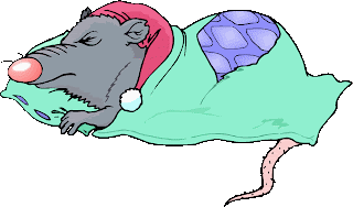

Even rats have to get some sleep. It was nice to have the sleeping cap,
but unnecessary for a sleep deprivation study. Not a good use of
research dollars.
“I’m dying for a good night’s sleep.” Is this just hyperbole, or an impending warning of death? For laboratory rats, sleep deprivation does kill. During their insomniac downward spiral, the rats tend to get hot and can’t cool down – you know, they can't thermoregulate (see Can’t We Just Go With The Flow). This doesn’t mean that a loss of the ability to thermoregulate is what kills the rats, but it does suggest a connection between sleep deprivation and the hypothalamus.

We looked at the hypothalamus in our story of endothermy. This evolutionarily old brain structure implements a set point temperature for the body and receives information about the temperature of different parts of the body. When the body temperature deviates from the set point, the hypothalamus initiates bodily mechanisms to normalize the temperature.


Apparently one of the effects of sleep deprivation is that you
become semi-transparent.
People with severe insomnia tend to sweat more and have higher core temperatures even though they say they are cold. They also have extreme high blood pressure, pulse, and appetite. These symptoms suggest that sleep deprivation messes with the hypothalamus, since functions of the hypothalamus include themoregulation, sleep, hunger, thirst, reproductive readiness in females, and stress responses. What scientists don’t know yet is just how sleep deprivation actually kills the rats or harms people.

Dying from a lack of sleep is not just a rat problem, a few very unlucky humans die from it as well. Fatal familial insomnia (FFI) is a very rare genetic disorder; it has been reported in only 40 families worldwide. Before describing the truly horrible way these patients die, let’s look at what causes the disease.

FFI is caused by a point mutation in the gene for the prion protein PrPc. A point mutation means that one nucleotide on the DNA is changed, which leads to a change in the protein coded for by the DNA. Three unit (nucleotides) segments of the RNA (made from the DNA template) work together (called a codon) to code for one protein building block (amino acid). In the case of FFI, the amino acid called aspartic acid is changed to one called asparagine, and this changes the protein’s shape. 


The left image shows mRNA bases recognized in sets of three
(codons) by tRNAs with amino acids attached (Ser = serine, tyr =
tyrosine). The amino acids are linked to because proteins. The
lower section is the genetic code, showing which amino acids are
coded for by which codons. The right image shows how proteins
fold. The primary structure is the amino acid sequence. The
secondary structure comes from interactions of adjacent amino acids,
including spirals called helices or sheets. The tertiary structure comes
from the folding up of the entire protein, while the quaternary
structure comes from the interaction of different proteins into a
larger complex.
PrPc is made up of 250 amino acids linked together in a chain. Each different amino acid carries a different shape and charge and will interact with every other amino acid differently. The sequence of amino acids in a protein cause it to fold into a specific shape. It is the protein’s conformation (shape) that determines its function. This is the opposite of what we determined for evolved organism characteristics, where form follows function (see Do You Have To Be Ugly To Hear Well?). With proteins – function follows form!

Mutation of that single amino acid at position 178 (aspartic acid is negatively charged, while asparagine is positive) causes the folding, and therefore the function, of the protein to change. Aspartic acid is sometimes abbreviated "D", while asparagine is called "N"; therefore, the mutation is often indicated as D178N (D at position 178 is changed to N).

Many genetic mutations result in no change in amino acid, or a change that bring a large enough change the shape to cause a change in function. But when it does, good or bad things can happen. On one hand, the altered protein might confer an advantage to the organism, one that promotes survival in the environment or after an environmental change.This positive selection through reproductive advantage become the new normal – and this is evolution

On the other hand, the change in amino acid sequence, form, and function could be destructive. Disease might be the result, or perhaps a change in the organism that reduces reproductive success. One of these two results is what occurs with the FFI mutation of the prion protein.

When the mutated prion folds differently, it forgets its day job and moonlights as a sinister evil force. Every other prion protein it contacts, WHETHER MUTATED OR NOT, is coaxed into changing its shape. The new prions turn to the dark side, then change other prion proteins they contact, multiplying the effect. The poorly folded prion proteins will stick together, come out of solution, and form solids (plaques) where they settle out. In different prion protein diseases, this settling out occurs in different parts of the brain. In FFI, it is the hypothalamus.


In the top image, the PrPc on the left is properly folded. The green
represents alpha helices and the blue arrows represent beta-pleated
sheets. The right image shows the malfolded version of PrPsc. It is a
tighter structure, which partially explains why protein-degrading
enzymes don’t work on it. . The lower cartoon shows that the PrPsc
can force the PrPc to assume the improper form, and these then
aggregate into plaques.
The prion plaques are longer lived then the regular prion protein; normal cellular enzymes whose job it is to degrade proteins won’t work on prion plaques. And worse, if some of the malfolded protein is transferred to another animal, the recipient will develop plaques and disease as well. That makes this an infectious disease that isn’t caused by a bacteria, fungus, parasite, or virus. The prion is an infectious protein! What a terrible exception to the rules of infectious diseases.

We see here a protein that can replicate itself (not by building more of themselves, but by changing the form of normal proteins), and that makes it a repository of biologic information. This is an exception to the central dogma of molecular biology, which says that DNA is the sole information storing material.

FFI moves from person to person through heredity, but if a non-affected person comes into contact with some brain material from an FFI patient and that material entered their bloodstream, it can be transmitted this way as well. A prion protein disease called Kuru is famous for being transmitted from person to person.

The Fore tribe in Papua New Guinea once observed a ritual wherein they honored a dead tribe member by eating part of their brain (called ritualistic mortuary cannibalism - gasp!). Because of this, there was an epidemic of Kuru in this tribe in the early 1900’s. Over a period of 3-6 months victims would become unsteady, irrational with bouts of laughter, and then degrade mentally and physically to the point of death. There are more than twenty known prion diseases (mad cow disease, Creutzfeldt-Jakob, scrapie, etc.), and Kuru suggests that some might have no genetic component, only person to person transmission.


A member of the Fore tribe is shown on the left. This tribe used
to celebrate the lives of departed members by eating their brains.
This spread a prion protein disease called Kuru, a protein disease
that is infectious! The Fore tribe still lives in Papua New Guinea,
although there are fewer of them than before Kuru.
The differences between the various prion diseases are based on the specific prion protein mutation, what part of the brain is attacked, and how potent the prion is at refolding normal prion proteins. For instance, the D178N mutation in FFI also occurs in Creutzfeldt-Jakob Disease (CJD), but a normal polymorphism (an amino acid change that doesn’t change form or function) at position 129 determines the fate. If amino acid 129 is methionine, the the person gets FFI, if it is valine, then they get CJD. 

The families that suffer from FFI have the D178N mutation, and also pass on the polymorphism for methionine (M) at position 129. Even more gruesome, some cases of prion protein diseases can be sporadic, not associated with either an inherited mutation or transmission. The malfolded prion can very rarely arise out of nowhere in isolated individuals.

The mutated PrPc is passed on via inheritance. You get one copy of each chromosome from each of your parents, so for an individual gene, you might get two normal copies, 1 mutant copy and 1 normal copy, or 2 mutant copies. Some diseases require that you must inherit two mutant copies for symptoms to show (recessive), but other require only one mutant copy (dominant, it dominates the trait from the other parent).

FFI is autosomal dominant (not associated with the X or Y sex chromosomes), so the chance of getting a mutant copy and the disease if one parent has it is 1 in 2; these are bad odds. But, if everyone with FFI dies, then why is the disease still showing up in families. Remember that we said above that some genetic diseases can, but don't have to, affect reproductive success. Unfortunately for those with FFI, the symptoms appear in the victims’ fifties, after they have had children. Natural selection doesn’t eliminate FFI from the population because FFI doesn’t appear affect reproduction.

The first symptoms of FFI include sweating while feeling cold. Later, the ability to get a good night’s sleep is lost, followed closely by the inability to nap. As the disease progresses, there are panic attacks, phobias, and no sleep whatsoever. After 4-6 months, mental abilities start to degrade. In its final stages unresponsiveness precedes death. 

This is especially sad way to die, because during the majority of the disease course the patient is aware of everything going on. At least with middle to late Alzheimer’s disease the patient is blissfully unaware of their dementia.


For both the gross and microscopic images, the left example is from prion protein disease victim, while the right example is from a normal brain. The brains on the left show how great the loss of tissue can be in Creutzfeldt-Jakob disease. The microscopic image from the diseased brain shows the plaques and the resulting holes in the brain structure. The small gaps in the normal brain on the right are a result of shrinking of tissue after it was on the slide.
On autopsy, the hypothalmus of an FFI sufferer looks like it has been hit with a shotgun blast. Holes are present in the tissue, representing areas where neurons have been lost due to inflammation and triggered cell death. The affected area of the brain takes on a spongy appearance, so prion protein diseases are lumped together and called transmissable spongiform encephalopathies (encephalon = brain and pathy = disease). Unfortunately, there are no cure, treatments, or vaccines for any of these prion diseases.

It is the hypothalamus' control of sleep cycles and circadian rhythms that promotes survival in animals. But what about plants? They don’t have a hypothalamus. Can they suffer from loss of circadian activity? In a word – yes!  And this will be our starting point next time.


For more information or classroom activities on prion proteins, central dogma, infectious or genetic disease, the genetic code or protein structure, see:

Prion protein and diseases –

central dogma of molecular biology –

infectious disease –

genetic disease –

genetic code –

protein structure –
nwabr.org/sites/default/files/learn/bioinformatics/AdvL5.pdf
 

Wednesday, January 20, 2016

Pump Up Your Brain


Biology concepts – learning, memory, attention, concentration, hippocampus, neurotransmitters, neurotrophins, executive function, processing speed, exercise


Many people exercise because of how it makes them feel,
or just because they think it helps them think more
clearly - maybe by reducing stress. They will be happy to
know that exercise actually increases the power of your
brain, everything from learning, to memory, to attention,
to decision making speed.
Many years ago, my father told me the story of how he studied while in college. He would hit the books in a solitary, silent room and just cram until he couldn’t concentrate anymore. Then he would get up, go outside, and run laps around his dorm for a while. Then he would come back and start again. Study, run, repeat. Turns out, the running makes a true difference. Exercise can actually make you smarter!

In a study from 2011, researchers took overweight kids and had them start exercising. Those that had at least 30 minutes of physical activity each day showed increased hippocampus size, and significant improvement on a CAS planning test, an alternative to the standard IQ test.

Planning basically means that their executive function (planning, reasoning, and decision making skills) had improved markedly. They also performed much higher on a math test, even though no additional math instruction had been given.

Exercise has impacts on memory, learning, attention, concentration, and processing speed. So now we know what we are talking about when we say exercise helps learning. Oh – you won’t just take my word that exercising helps? Good, always ask for evidence.

Let’s look at studies just from 2013, although there are many older studies. One study found that a single bout of moderate exercise allowed participants to more accurately complete a test on memory, reason, and planning - and it took them less time. Another study indicated that exercise reduced the loss of cognitive function in middle-aged women. Yet another publication talked about how master athletes (over 50), have a larger brain volume and better cognitive function as compared to their sedentary counterparts.

We can go on. Exercise has been shown to support the cellular structure of the white matter (myelinated) neurons of the cerebral cortex in patients with vascular disease, important for higher thinking functions. And another study shows that processing speed is increased after starting a regular regimen of cardiovascular activity.


The upper image shows where the hippocampus is located
within the brain. There are two, one in each hemisphere.
They are connected as well. The lower image shows the
regions of the hippocampus, including the dentate gyrus
(DG) the area where much of the neurogenesis after
exercise is found.
Finally, we will mention just one of the many 2010 studies. Nine-ten year old kids that exercised regularly had 12% larger hippocampi (plural of hippocampus, part of brain for learning and memory). They were faster on recall tests and they learned new information faster.

So now that you are convinced that exercise does help cognitive functions, the question still remains as to how exercise carries out this miracle. The first thing to get clear is the difference between memory and learning. It might seem that they are the same thing; you have some experience, either verbal, aural, visual, etc. and if you remember it, then you have learned it. But there are subtle differences.

Specialists define learning as a process that will modify a subsequent behavior. Memory, on the other hand, is the ability to remember past experiences. Memory is the record left by a learning process, so you need to have memory to learn. You learn to play piano by studying the notes and the instrument, but you then play it by using your memory to retrieve the notes and fingering that you have learned.

Back to the mechanisms of how exercise help memory and learning. The easy explanation is that exercise helps you sleep, improves your mood, and drives more oxygen to the brain. These undoubtedly help you study better or even notice more that can be used to build knowledge. These are the factors my dad counted on when he went running. But there’s much more.

The hippocampus is important for learning and memory. Many studies of exercise and cognitive function have shown increases in the size of this part of the brain in exercise participants. Those kids that increased their “IQ,” they had an increased hippocampus. So did mice from studies in the 1990’s.


Neurogenesis is the production of new neural cells from
stem cells. There are stem cells located in the brain. They
can become any type of brain cell, depending on stimuli in
the local area. Normally, only a small percentage of
stimulated stem cells will become neurons, but after
exercise the number that survive goes up dramatically.
Exercise upregulates neurogenesis, oxygenation, synaptic plasticity, neurotransmitter populations, myelination, processing speed, and long-term potentiation (LTP). O.K., that’s a lot of big words, so let’s take them one at a time. Remember that all these things are linked together. Plasticity, neurogenesis, and LTP apply to memory. Neurogenesis and processing speed apply to new learning and executive function. Neurotransmitters, plasticity and oxygenation combine to affect attention.

Neurogenesis
A lot of the benefits from cardiovascular exercise come through the making of new neurons (neurogenesis). Yep, this is a huge exception to the rule that central nervous system neurons last your entire life and can’t be recovered or new ones produced. Neurogenesis is how the hippocampi of all those exercisers got bigger.

Regular exercise induces neurogenesis through action of brain chemicals, trophins and NTs. We talked about brain-derived neurotrophic factor (BDNF) 2 weeks ago with respect to mood and we said we revisit this factor. This neurotrophin actually stimulates your brain to make new neurons! More neurons means more connections, and more potential learning.

For most all of the cognitive functions, the lynchpin seems to be BDNF. How does exercise increase BDNF? We aren’t sure yet. It may be that exercise is a stress, this increases the calcium flowing into the brain. The calcium activates many transcription factors, and BDNF is known to require calcium for transcription.

But nothing is ever simple. It is probable that serotonin, IGF-1, and BDNF are all needed to increase neurogenesis in the hippocampus. Inhibitors of any one of these drastically reduce the amount of exercise-induced neurogenesis.

Plasticity
Think of plasticity as a general process, the altering of neurons and their connections. It involves making more neurons (neurogenesis) and the number (developmental plasticity or synaptogenesis) and orientation of the dendritic connections with other neurons (synaptic plasticity).


The images show the increase in the number of dendrites and
possible synaptic junctions over time. The increase in dendrites
is called arborization (arbor = tree) for obvious reasons. The
increase in synapses is called synaptogenesis. Exercise increases
both of these. This image isn’t a result of exercise, but it would
be is similar.
BDNF doesn’t just induce new neuron formation, it can increase the number and size of the connections (synapses) between neurons. IGF-1 is probably involved in this as well, as its main function is to support the growth of fragile, newly formed neurons and connections.

Plasticity is crucial to learning and to memory, since all learning and memory is just a map of connected circuits that work together to access certain information. It is the number and pattern of the connections that determine the amount retained. More connections must help this process.

Long term potentiation
LTP is for memory and learning – the reinforcing of neural connections to make them stronger. Exercise increases LTP, probably through synaptic plasticity, more connections between two neurons would help reinforce each other when they fire. We talked about LTP last year, so read that post and know that exercise increases it.

Unfortunately, for best increases in memory the exercise must be long term. In a 2013 study, neurogenesis was apparent only 14 days after initiation of exercise, and these were immature neurons. LTP wasn’t increased appreciably until 56 days.

Processing speed
Increased speed probably comes through increased IGF-1 and oxygenation, and their effects on the support cells in the brain. Oligodendrocytes and astrocytes help the neurons do their job at peak efficiency. In particular, oligodendrocytes make the myelin sheath that increases transmission speed.


Meet the glial cells. Astrocytes mediate the travel of fluids
and nutrients from the capillaries to the neurons and
between the neurons and the cerebrospinal fluid.
Oligodendrocytes make the myelin sheath around some
axons. Microglial cells are the immune system of the brain,
they phagocytose intruders. Finally, the ependymal cells
line the ventricles in the brain, the spaces that hold the
cerebrospinal fluid.
Astrocytes, on the other hand, are important for blood flow to neurons, and cerebral spinal fluid movement. These two functions would be particularly important for moving neurotrophic factors toward the neurons.

Attention and concentration
Exercise also helps your attention and concentration. And no, these two aren’t exactly the same. They’re more closely related than memory is to learning, but there are still some differences. Both are important for making you smarter, because only by focusing do we take information in – you have to notice something to learn it.

When you are in a room full of people talking, you can still follow the conversation between yourself and one other person. This is one of several different forms of attention. In general, attention is a thinking process for directing and maintaining awareness of stimuli in one’s environment.

Concentration is the ability to control attention for a sustained period. Attention shifts as we wander from thought to thought about different things in our mind or environment, but concentration requires attention to one thing without wandering. In more clinical terms, concentration is a combination of two types of attention; sustained attention and selective attention.

Sustained attention is staying on task, keeping your mind on a single task over time. Selective attention is more about how you pick what you pay attention to. If there are many activities going on within range of your sense, but you focus on one thing and pay no attention to the others, that is selective attention.


Attention span is not equal to sustained attention. It is
focused attention; how long until your brain diverts to
some other stimulus. In 2000, Americans had a 12 second
attention span on average. In 2010, it was down to 8
seconds. Heck, a goldfish has a 9 second attention span,
and we make fun of them!
Attention is centered in the reticular activating system (RAS), near the brain stem. But it connects to other centers that work in attention as well, like the prefrontal cortex and the parietal cortex. The RAS accounts for shifts in levels of awareness to different things. Exercise activates the RAS, which increases alertness, and therefore attention and concentration – my dad was ahead of his time.

It turns out that increased dopamine, serotonin, and norepinephrine in the brain, and particularly the RAS is crucial for attention and concentration. And we talked two weeks ago about how exercise increases all these. In ADHD, they give drugs (methylphenidate) that increase the apparent levels of dopamine. This helps us make sense of studies that show regular exercise alleviates the symptoms of ADD/ADHD.

One last point that I find interesting. The type of exercise seems to make a difference for the increase in neurotrophins. A 2012 study showed that rats that ran on exercise wheels had increased BDNF in the hippocampus, but rats that lifted weights (climbed ladders with weights on their tails) increased only IGF-1. The two proteins work in different pathways, so rat studies show us that it is best to include both aerobic and resistance training in your exercise program. And a rat shall lead them.

Next week, can you die from not getting enough sleep. Yep, and that's not the weirdest part of fatal familial insomnia.


For a good resource on brain structure and function, see the Open College’s interactive brain.


Patten AR, Sickmann H, Hryciw BN, Kucharsky T, Parton R, Kernick A, & Christie BR (2013). Long-term exercise is needed to enhance synaptic plasticity in the hippocampus. Learning & memory (Cold Spring Harbor, N.Y.), 20 (11), 642-7 PMID: 24131795

Cassilhas RC, Lee KS, Fernandes J, Oliveira MG, Tufik S, Meeusen R, & de Mello MT (2012). Spatial memory is improved by aerobic and resistance exercise through divergent molecular mechanisms. Neuroscience, 202, 309-17 PMID: 22155655

Davis CL, Tomporowski PD, McDowell JE, Austin BP, Miller PH, Yanasak NE, Allison JD, & Naglieri JA (2011). Exercise improves executive function and achievement and alters brain activation in overweight children: a randomized, controlled trial. Health psychology : official journal of the Division of Health Psychology, American Psychological Association, 30 (1), 91-8 PMID: 21299297

Tam ND (2013). Improvement of Processing Speed in Executive Function Immediately following an Increase in Cardiovascular Activity. Cardiovascular psychiatry and neurology, 2013 PMID: 24187613

For more information or classroom activities, see:

Most of the information for this post comes from recent scientific journals, here is more general information from the internet.

Memory classroom activities –

Hippocampus –

BDNF –

Neuroglia –

Wednesday, January 13, 2016

Exercise Puts Me To Sleep – You Too

Biology concepts – sleep induction, circadian cycle, narcolepsy, insomnia, anterior hypothalamus, neurotransmitters, cytokines, inflammation


Harriet Tubman gained the respect of all after the Civil
War, including that of William Seward, Secretary of State
of the United States (he’s the guy that bought Alaska).
Despite this respect, she ended up penniless. Seward
provided her with a two story brick house in Auburn,
New York where she could live out her days in
relative comfort.
Harriet Tubman was a narcoleptic. No, she didn’t steal things from the Woolworth, that’s kleptomania. She led hundreds of slave to freedom in the years before the American Civil War despite fighting off the urge to go to sleep at any given moment.

Narcolepsy is a sleep disorder that affects 1 in 2000 Americans, and causes them to have episodes of extreme fatigue. They fall asleep at odd times, and are very hard to awaken. In addition, they may also suffer from wakeful dreams, and cataplexy, a condition similar to temporary paralysis. This can’t have instilled confidence in Harriet’s passengers, but she got the job done.

Narcolepsy is basically too much sleep induction, while insomnia is too little. Many people suffer from insomnia, and it can be brought on by many different conditions. Could your New Year’s resolution to start exercising end up helping both narcolepsy and insomnia. Let’s find out.

You wake up in the morning determined to wear yourself out on the treadmill sometime today. You’re going to run to exhaustion in the hopes that you will get a good night’s sleep as a result. Your doctor did say that working out would help you sleep – but is this what he meant? You run until you’re out of energy and then you sleep to refill the gas tank?

Exhaustion from exercise may play a role in inducing sleep, but there’s much more. Exercise helps in insomnia in the elderly according to some reports. Exercise also helps with narcolepsy in children and adults. But how does it help?

There are two competing hypotheses for exercise’s effects on sleep via your brain. They use two different sensors, but they both run through the brain. But first we need to know a little bit more about the sleep center of the brain to explain how exercise is affecting us.

Sleep was the subject of a series of posts a couple of years ago, starting with this post. But we didn’t talk much about the induction of sleep. Sleep used to be considered a passive process; you slept when the stimulatory inputs to the brain were diminished.


Here is the hypothalamus, home of the sleep centers of the
brain. One the left you can see where the hypothalamus is
relative to the rest of the brain structures. On the right is a
cartoon showing the hypothalamus closer, including the
VLPO, the SCN, the LHA for wakefulness, and the MPO
for heat regulation.
Sleep is now known to be an active process, controlled by the anterior hypothalamus and preoptic nucleus. See the picture to help you locate this area of the brain. In the anterior hypothalamus and the preoptic nucleus, stimulation of GABAergic neurons promote sleep induction and maintenance. GABA is a neurotransmitter that is inhibitory, it stops some of neurons from firing. In this case, the neurons it inhibits are the ones that produce orexin/hypocretin. This is another neurotransmitter, but this one stimulates wakefulness.

Orexin is one neurotransmitter with two names. It was discovered by two different groups at just about the same time, and each group named it something different. Scientists haven’t decided yet which name to go with, so they use both.

There are only 10,000-20,000 neurons which produce orexin/hypocretin, so damage to any part of this area of the brain could induce narcolepsy. This may have been what happened to Harriet Tubman after her master hit her in the head when she was 12 years old. On the other hand, the brain trauma may have resulted in too much VLPO and AH sleep promotion. Either way, she was a professional napper.

So stimulating the POAH and the VLPO lead to sleep at least in part by inhibiting the production of orexin/hypocretin. But what stimulates the POAH and VLPO? Knowing nature as you do, you can bet there are several pathways. One way is certainly routed through the circadian clock. We have talked before about the sleep cycle controlled by the clock.


Yet another picture of the brain, this time highlighting the
pineal gland, where melatonin is made. The left side suggests
that light affects the pineal, but it ain’t the way the yellow
arrow shows. The right figure shows the true pathway much
more realistically. The pineolocyte is the cell type found in the
pineal gland. The melatonin is made from tryptophan and
serotonin is an intermediate structure, so you can make it
from serotonin itself.
Different hormones (like melatonin) and neural inputs/outputs stimulate the sleep and wake centers of the brain to create a semi-regular day/night cycle. Many things can mess with the cycle of melatonin and other day/night rhythms, including exercise. Now we can talk about different ways this may occur.

Temperature hypothesis:
The temperature hypothesis for sleep induction states that a one-degree decrease in your core temperature is enough to trigger sleep induction pathways on the brain. How could these two factors be linked? Well, the temperature sensing and regulating centers of your brain are located in the anterior hypothalamus, right next to the sleep centers (POAH and VLPO).

Reducing temperature is a way of saving energy by the body; this is probably an evolutionary holdover from when calories were hard to come by. Decreasing temperature signaled the brain that less activity was going on, so the body induced sleep to further reduce temperature and save energy for the next day.

It so happens that activity also decreases when the sun goes down, or at least it did before Thomas Edison and the electric light. This strengthened the link between temperature and the circadian sleep/wake cycle. To illustrate this point, a 2013 study measured the effects of drugs on both the circadian patterns and temperature. Drugs that altered the light responses in the SCN, including caffeine, also altered core temperature.


GABA (Gamma-aminobutyric acid) is a neurotransmitter
release at the synaptic cleft of some neurons. It is inhibitory
for some wakefulness neurons, so it promotes sleep. On the
other hand, noradrenaline is stimulatory for the orexin
producing neurons so it promotes being awake. In the middle
is adenosine, you know the player in DNA, RNA, and ATP. It
also happens to be a neuromodulatory and can inhibit the
GABA, NA, and orexin neurons.
In the same study, the same responses that reinforced circadian cycle (spontaneous sleep about 16 hours after light stimulation) also reduced the core temperature at the same time. Drugs that inhibited one, stopped the other as well. It would appear that temperature and day/night cycles are very much linked for sleep.

That then brings up the question of how exercise helps you go to sleep just by messing with your temperature. You exercise - you get hot - the blood vessels in your skin dilate and you sweat to dissipate some of the heat. But sweating isn’t 100% effective, your core temperature does go up. After you finish exercising, your temperature goes down slowly over time.

This decrease in temperature is the cue for your body to begin sleep. Your anterior hypothalamic temperature-regulating center can’t tell the difference between this decrease and the decrease brought about by circadian rhythms. So you may get sleepy a few hours after exercising, as your temperature comes down.

So, is right before bed the best time to exercise? Nope. Exercising stimulates your brain and cardiovascular system as well as raises your temperature. Trying to sleep right after exercise will probably be harder than normal, just because you are firing on all cylinders in your brain and heart.

The best time to exercise to help you get to sleep is about five hours or so before you plan on retiring for the evening. Your temperature goes up while exercising, and then will start to drop just about the same time you are ready for bed. This will reinforce the circadian cycles and give you the best shot at good sleep.


Insomnia is found in a number of conditions. It is
becoming a serious problem in the elderly, with people
living longer and unfortunately becoming more sedentary.
Complications are shown in the cartoon. You see that the
effects are varied, including the ability to fight off
disease. Who knew that not sleeping could lead
to diabetes!
The above plan applies to most of us, but perhaps not all of us. Very well-trained athletes might be less affected by the changes in body temperature for sleep induction. One 2013 study looked at exercise time, temperature manipulation and sleep patterns in professional and highly trained amateur cyclists. The results showed that evening exercise had no affect on sleep patterns, even if combined with a cold water dunk after the cycling routine (brrr!). Neither exercise nor exercise + decreasing temperature brought on a decreased time to spontaneous sleep. So – they sleep well because they wear themselves out each day.

Cytokine hypothesis:
The other system that may be important for inducing sleep after exercise is the immune system. Cytokines are chemical messages that influence many different parts of the immune system. They come into play when you have an infection, or cancer, or allergy; basically any insult to your system.

There are many different cytokines, and they perform many different jobs. Certain cytokines can even mediate opposing pathways, depending on the stimulus that starts their production and release. Some promote inflammation (pro-inflammatory) when one specific injury is sense, but inhibit inflammation (anti-inflammatory) if a different insult occurs.

Exercise can be seen as a stress to the body. It can injure muscles; in fact, that's how you build muscle. You tear them down a bit through work, and they grow back bigger and stronger. This is an insult that results in cytokine production and release into the bloodstream. But the more you train, the less of an insult your body registers.

IL-1beta, TNF-alpha and IL-10 are cytokines that have been associated with sleep induction. Plasma levels of IL-1beta are highest just as sleep is induced; this is one of the things controlled by the circadian system. But prolonged IL-1beta or TNF-alpha results in short sleep, and it is easy to wake you up.


Cytokines have big roles in the brain, even if they
act indirectly. Second messengers trigger pro-
inflammatory cytokines through the brainstem
which control fever and other symptoms,
including sleepiness. You think it’s a coincidence
that you sleep more when you’re sick?
On the other hand, IL-10 is anti-inflammatory and is higher with physical training over time. A 2012 study showed that in the elderly with insomnia, moderate training over months resulted in lower IL-1beta, lower TNF-a, and higher IL-10. These were also associated with better sleep patterns.

The neurons of the sleep center are sensitive to pro-inflammatory cytokines; inflammation signals disrupt the restful sleep patterns we are looking for. This involves the pro-inflammatory stimulation of cortisol, the stress hormone, so exercise’s help in sleep may be again related to a reduction of stress effects. This is a complicated system, but the take home message is, more exercise results in less pro-inflammatory cytokine action on the brain.

Chronic fatigue is also linked to high levels of pro-inflammatory cytokines in the brain that are unhooked from the decrease induced by training for some time. In the opposite direction, narcolepsy is aided by exercise, perhaps by reducing the cytokines that would inhibit orexin/hypocretin domination (wakefulness) in the anterior hypothalamus.

A 2007 study showed that in mice that don’t make orexin/hypocretin (have narcoplepsy), running on the wheel helped them stay awake more during the day. Of course, it also led to more episodes of cataplexy, so the story is not complete.

Next week, another brain effect of exercise – it can actually build your brain and make you smarter. Start running before that next AP quiz.


For a good resource on the structures of the brain, see Open College's Interactive Brain map.


Vivanco P, Studholme KM, & Morin LP (2013). Drugs that prevent mouse sleep also block light-induced locomotor suppression, circadian rhythm phase shifts and the drop in core temperature. Neuroscience, 254, 98-109 PMID: 24056197

Robey E, Dawson B, Halson S, Gregson W, King S, Goodman C, & Eastwood P (2013). Effect of evening postexercise cold water immersion on subsequent sleep. Medicine and science in sports and exercise, 45 (7), 1394-402 PMID: 23377833

Santos RV, Viana VA, Boscolo RA, Marques VG, Santana MG, Lira FS, Tufik S, & de Mello MT (2012). Moderate exercise training modulates cytokine profile and sleep in elderly people. Cytokine, 60 (3), 731-5 PMID: 22917967

EspaƱa RA, McCormack SL, Mochizuki T, & Scammell TE (2007). Running promotes wakefulness and increases cataplexy in orexin knockout mice. Sleep, 30 (11), 1417-25 PMID: 18041476
For more information or classroom activities, see:


Exercise and sleep –

Narcolepsy –

Orexin/hypocretin –

VLPO and sleep –