Showing posts with label nature of science. Show all posts

Wednesday, July 27, 2016

The Nature Of Science Of Nature

I have judged many a science fair project in my
day. It is a learning experience, so adherence to
the steps of the scientific method taught in most
classes is O.K. But the students should realize
that there isn’t just one scientific method. This
young lady’s strategy was to take all the available
time and be the only project that got judged;
therefore, she's the winner.
A summary of the usual (but hopefully disappearing) lecture on the scientific method –
      1.     Define a problem
      2.     Research what is known
      3.     Hypothesize a cause
      4.     Test your hypothesis
      5.     Analyze your results
      6.     Support or refute your hypothesis

In the next few class periods, a few examples are identified and the class works through them. And there is always the "design an experiment to prove your hypothesis."

The question of the day:
Is this the only method to conduct science?

I don’t have room to go into all the things that are wrong with the process as it is enumerated above, but let’s hit a couple.

One big problem is the idea of supporting or refuting your hypothesis (hypo = under, and thesis = proposition; a supporting basis for an argument). True, your experiment may do one or the other, but it doesn’t have to. Many experiments end up with ambiguous results, especially if the scientist is doing his or her job.

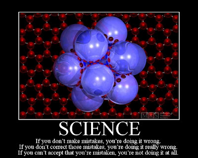
I am using this picture because I couldn’t find a
way to work the quote into the post. But in truth,
science is like life, better results often come from
many trials. So... science is like life which is like 
science.
The design of the experiment should minimize any confounding effects that would render the results less than explicit. But, as the old saying goes, “You don’t know what you don’t know.” The middle of an experiment is often a learning moment. It is when you get into a study that you find the things that are going to make the study useless. Then you go back and design a better experiment.

Let’s say you have designed an experiment that will have a definitive result. What result are you going for? Most will say that you are trying to see if your hypothesis is supported. But anyone can design an experiment to give a desired result. Your hypothesis says “A” should occur if you do “B,” so you design an experiment to make sure “A” occurs. It doesn’t even have to be a conscious effort, your design will just tend toward that result.

A truly scientific experiment is designed to REFUTE your hypothesis. You design an experiment to prove your hypothesis is not true. If, under those, conditions, your hypothesis is still supported, then you really have something. If a scientist tries his/her best to refute their own hypothesis and can’t, the chances that the hypothesis is correct go way up. Then you report your results and let other scientists try to design an experiment to show your hypothesis is wrong. If they can’t do it either, then you are really onto something. Real scientists try to prove themselves wrong, not right. The big idea - you can never PROVE a hypothesis, you just have data to support it. But the next experiment may refute it. You can always design another experiment to test the hypothesis, so no hypothesis is ever proven absolutely. True science proceeds when you refute a hypothesis; only then can you make a concrete change to move closer to the truth.

Negative results and refuted hypotheses are the basis of science; 
too bad they get a bad rap.  Can you think of another profession 
where being wrong is your goal?
This leads to the next problem implied by the process outlined above. It is usually taught that negative data is a bad thing. Even people in the profession often downplay the importance of negative data; ie. data that does not give a result that is publishable.

Editors don't get excited over studies saying we tried this and it didn't work. But, an experiment that doesn’t work isn’t necessarily bad – you can learn a lot from it and so can other scientists. Unfortunately, journals don’t like to publish this data, so those who might learn from it don’t get to hear it.

This is one reason why it is important for scientists to have meetings and talk to one another personally, not to just write journal manuscripts and funding applications. Case in point, it turns out that studies that show new drugs aren't cure-alls, that they don't do what they say or don't do it as well as they say don't published. Ben Goldacre gave a recent TED talk on the subject, and has numbers to back up his assertions that negative data studies on drug efficacies hardly ever see the light of day.

In fact, negative data is the most common data and often the most useful. Refuting your hypothesis is a type of negative data. When faced with this result, you modify or discard your hypothesis and try again. You can design a thousand experiments that support your hypothesis and still not prove that your idea is the true mechanism - you may just not have thought of the experiment to disprove it yet. Like we discussed above, just one experiment that DISPROVES your hypothesis results in a step forward. Like Thomas Edison said, "If I find 1000 ways something won't work, I haven't failed. I am not discouraged, because every wrong attempt discarded is another step forward."

Negative data truly moves us forward, in fact failing is the only way we move forward. But this still leaves a problem with the way science is taught. Is there good data that is neither positive or negative? We tend to think of data only as that information that supports or refutes a hypothesis, but do you have to have a hypothesis?

This is the black walnut tree (Juglans nigrans).
The one in our front yard is about 70 feet tall and
produces over 200-300 kg (500-650 lb) of fruit.  
Black walnut dye comes from the husk, not the 
nut, and is yellow when immature and black 
when mature.
Consider an experiment I have been conducting for the last 13 years. We have a black walnut tree in our front yard, and I have been counting the number of black walnuts it drops every year since we moved in. I had no mechanism I was trying to define, I just wanted to know how many walnuts the tree produced.

Here is my data:
Year                        # of walnuts
2003                        3662
2004                        604
2005                        3508
2006                        368
2007                        4917
2008                        0                           
2009                        6265         
2010                        0             
2011                        6395
2012                        6
                                                                                       2013                        2140
                                                                                       2014                        159
                                                                                       2015                        1825

Now, we can ask a question and hypothesize a mechanism? What is responsible for the pattern in nut production, and why do the results keep diverging? Does that mean that my original observations aren’t science? True – you could say that I was answering a question about how many nuts the tree produces, but I did not have a hypothesis that I was trying to dispute. This is true science, but not the kind we teach in school. Is the change in the pattern during the middle years accounted for by weather? Do the fewer nuts later mean that the tree is dying?

Black walnut meats are expensive because
they are hard to get out of the shell. But the
shell is also economically important, used in
paints, oil wells, explosives, cosmetics, cleaning
and polishing agents, and jet blasting of metallic
and plastic surfaces.
Try this experiment at your school. Find a tree and start to count the nuts, or find a sapling and count the leaves each year. You can keep this experiment going over a number of years, with each class adding their data.

But the real learning is in defining the limits and possible confounding effects that could lead to errors. Did the tree lose a limb and therefore produce fewer nuts the next year? Was there an explosion in the squirrel population, and they stole all the nuts before you could count them? What was the weather over the time period you observed, could a change in weather account for a change in number? Is there another walnut tree too close, and the nuts are getting mixed up? Am I just getting better at finding and counting the nuts each year?

Squirrels are kind cute, if you forgive them for
carrying rabies. Here, he represents one source
of possible error in my black walnut counts. Can
you assume that every year they steal about the
same percentage of nuts before I can count them?
I have asked these questions and am observing multiple parameters to see if they account for the pattern in nut production. But there may also be a biologic reason, something to do with energy output versus opportunity to produce offspring. All these items can be investigated and used to better explain the observations.

Each might be considered a hypothesis – the weather affects nut production, so you try to show that different weather years had the same nut production – hypothesis refuted. The squirrel population exploded – talk to the local nature experts, if the number has been fairly constant- hypothesis refuted. There is an almost infinite number of possible confounding effects, and your class can come up and test as many as their brains can think up. Now that is a true scientific method!


Ben Goldacre (2012). What doctors don't know about the drugs they prescribe. TED MED 2012

Wednesday, September 3, 2014

Bacteria Are Intelligent Designers

Biology concepts – nature of science, flagella, intelligent design, irreducible complexity, motility, Gram+, Gram -, ion gradient

You don’t believe it now, but in the weeks ahead we’re going to discuss how bacterial motility, plant reproduction, intelligence, and the location of your heart are all related to whips and eyelashes. Sounds preposterous, but give me a few posts and a little leeway and you’ll be amazed.


Cheetahs can cover about 25 body lengths in a second, but
some Salmonella can move 60-80 of their own lengths in the
same time! See this post for finding out what the fastest
organisms are. Salmonella typhi is the bacterium that causes
typhoid fever and is spread in contaminated water or touch.
Mary Mallon was blamed for 51 cases of typhoid fever as a
carrier (no symptoms but still sheds bacteria). A 2013 study
shows that the bacteria turn on a fat regulator, PPAR-delta, in
macrophages which lets them live inside the cells forever.
Let’s get right to it. Bacteria are small, but they’re quick little devils. They have inboard motors – or are they outboard? – I can never keep those straight. This piece of machinery is so complex and fascinating that some people use it as a sign that someone or something had a hand in designing life on Earth.

The bacterial motor is called the flagellum, but it's so much more than just a way to get around, it’s often the means to saving their own lives. The word flagellum comes from the Latin word flagrum meaning whip, so you can see we are already starting to work on our challenge for these posts. Flagrum could also mean scourge, and this seems to be prophetic, since many flagella (the plural) we study have a hand in causing disease.

In typhoid fever, a potentially deadly disease that affects more than 20 million people each year, flagella are important not just for putting the bacterium, Salmonella enterica typhi, in the correct place to cause disease, but for attaching the bacterium to the gut wall and for invasion of the gut. A study in 1984 showed that even flagella that couldn’t move were still needed for S. typhi to produce disease. More about this in a couple of weeks.

A flagellum is very much like a boat propeller, it spins to produce force along its axis. This is possible because flagella aren’t perfectly straight. One of the main components of the flagellum is called the hook. This hook is located just outside the cell wall and lies in between the basal body and the filament. The basal body is the engine and is what attaches the flagellum to the cell, while the filament is the long whip like end that sticks out into the world.


A cartoon of the bacterial flagellum shows the structures,
filament, hook, and the entire lower part is the basal body
(motor). The filament is hollow and sends new flagellin
subunits up through it to be added on to the end. The electron
microscope image shows the basal body. It looks like art or
engineering.
The hook turns at about 90 degrees, but the degree of turn is different for different bacteria. This means that that filament, when spun by the motor in the basal body whips around in a circle, bigger at the bottom where the hook is located. So instead of rotating like a straight pencil and not generating any forward force, it spins like a propeller.

The basal body attaches the filament and hook to the cell, and is made up of several rings. In Gram+ bacteria there are two basal body rings that anchor the flagella apparatus, the M ring which attaches to the membrane and the P ring which is anchored in the peptidoglycan layer. In Gram- bacteria, the basal body is longer and has more rings since it must anchor the flagella into the LPS (the L ring) and the M ring has a buddy in the inner membrane called the S ring. All these rings support the rod, which is turned by the rotor and then spins the hook and the filament.

The filament is pretty cool. It’s either a left- or right-handed helix of subunits of a protein called flagellin. The filament is a prescribed length in each bacterium, but we aren’t exactly sure how the length is regulated. Scientists know that it grows faster at first and then slows down, but if broken it will start to grow again at the faster pace.


The filament of the bacterial flagella is capped by a small
protein called FLiD. This is an amazing protein that
regulates and mediates the assembly of the filament
subunits of flagellin at the tip of the growing filament.
The flagellin units are straight as they travel through the
middle of the filament, but their final shape is bent. The
FliD mediates this folding at the tip.
The amazing thing is that the filament grows from the tip, not the base where it attaches to the hook. The flagellin filament is hollow, and subunits of the protein travel from the cytoplasm up through the basal body and hook and then through the existing filament out to the end. Then they are attached to make the filament longer. That’s a pretty neat system because it alleviates the need for a way of exporting the parts, regulating their movement to the end of the filament and then attaching them. Sometimes, but only sometimes, evolution finds the simpler way to do something.

The energy for the motion of the flagellum comes from the movement of ions across the membrane of the cell. We have seen before how protons (or other ions) being pumped out and then allowed to enter through a pore can create the force needed to do work. That’s how ATP is made, how the neural action potential works, and how photosynthesis proceeds. But here, the proton motive force is used to spin the hook and the filament, driving the bacterium forward.

The flagella spin one way to move forward, but when they spin the other way, the bacterium just sort of tumbles around. We’ll talk more about this next time. We’re just now starting to learn how the motor can go from spinning counter clockwise (forward motion for a left handed filament) to spinning clockwise in no time whatsoever and without slowing down. Nothing looks very different in the basal body, the hook or the filament, but the direction of spin is reversed.


This is a complicated picture so stick with me. A) is the shape
of the FLiG protein from a certain bacterium. The end we are
interested in is red, it holds the charge for interacting with the
ion gradient across the membrane. B) shows the positive and
negative bubbles of charge in the helix. Below, see the ring of
FLiG proteins of the rotor. When spinning different directions,
the positive and negative bubbles are reversed, one shape
makes it go clockwise, the other, counterclockwise. It all has to
do with the pushing and pulling by same and opposite charges
as the ions pass through the membrane.
I can hear you thinking out there, “Well, just reverse the direction that the protons move, instead of outside to inside, go inside to outside.” Nope, when a flagellum switches direction, the protons keep moving the same direction. We do have information that one of the proteins that connect the motor (electrochemical gradient) to the physical turning (rotor), a protein called FLiG, can change shape.

Several studies have shown this change, and it is hypothesized that the change moves charged amino acids of FLiG around in relation to the cation gradient. By changing them, it changes the direction of the turning of the rotor (see the picture to the right). This might be akin to reversing the poles of a mag-lev train by flipping the electrical charge can make the train go the opposite direction.

Different bacteria have flagella that look similar but they have small differences. Nevertheless, it can be seen that this is a very complex machine for such a supposedly “primitive” domain of organisms. We have to remember that bacteria have been here the longest; they must be doing something right. There are over 40 genes that are required to build a flagellum, and they all fit together just so.

This complexity and order leads some people to declare that flagella couldn’t have evolved on their own. The concept is called irreducible complexity. People who support the idea of intelligent design (ID) say that some biologic components are so complex and have so many working parts that they could not arise through a series of mutations.

All the parts of a flagellum must be present for it to work (therefore they say it is irreducible) and must be assembled all at once which suggests it could not be random (complex in ID means improbably occurs by chance). Therefore, a flagellum could not have evolved over time and, ipso facto, it must have been designed as one unit by someone or something.

ID proponents haven’t always focused on the flagellum. They first talked of the blood coagulation cascade as irreducibly complex, but then it was shown that portions of the cascade were not necessary for function – whales don’t have factor XII and jawless fishes only use about half the proteins that vertebrates use. It was also shown how the cascade evolved over time.


A vibrio bacterium can make two different flagella types,
signified by the two sides of the dotted line above. The ions are
different that run the gradient, and the genes are different for
the motor/rotor. Are there two different irreducibly complex
flagella or did one modify into the other – then they aren’t
“complex.” Vibrio vulnificus is shown on the bottom. It has been
unusually numerous this summer (2014) and causes a disease
that looks like flesh eating disease, but isn’t.
Over the years, ID has proposed that the eye, the immune system, the flagellum and the eukaryotic cilia and its production system were irreducibly complex.  But each time, the ideas of specified, irreducible and complex (must have all come together at once) have been refuted for each example.

For the bacterial flagellum, arguments against ID include the facts that different bacteria use different systems, although they are all variations on a theme. One exception is the Vibrio. They use two different kinds of flagella on the same cells, each needing its own genes. Likewise some bacteria don’t use protons for the gradients, they use Na+ ions. The bacterium Vibrio parahemolyticus is an exception in both cases.

It uses a single flagellum at its end (polar) to swim in liquid water, but many flagella all around its cell when in something thicker. The polar flagellum uses Na+ ions to drive the rotor, while the lateral ones use protons. The genes are different for each flagellar type and mutations in one don’t hurt the other.


The top cartoon shows that when a gene duplicates (and they
do, often) one copy can drift and acquire mutations without
hurting the cell. This can lead to better function or new
function. Over generations, one set of genes for a function
can be replaced with another set – this would hardly be
called irreducibly complex. On the bottom, you see the type III
secretory system for injecting bacterial toxins on the left and
the flagellum on the right. They are very similar, so why is the
flagellum irreducibly complex and the not the type III system?
Lastly, only some Vibrio and other bacteria have a protein sheath over their flagellar filaments. These protein sheaths cover the filament and aid in sensing changes in chemicals outside the cell. So which flagellar type is irreducibly complex and which is not?

Spirochetes don’t even have flagella that protrude from the cell, they’re located between the inner and out membranes (endoflagella). This is a different system and again argues against irreducible complexity in flagella, unless different systems were designed differently. More about spirochete motion next week.

Please read more about ID and decide for yourself if it holds up to the tenets of science - that something that is true must be observable, repeatable, and able to be refuted if incorrect. Irreducible complexity is refutable, and has been for every example proffered by ID. But the conclusion that ID draws – that a designer must be involved, is a belief not a hypothesis – you can’t refute a belief, it doesn’t rely on observable evidence, therefore ID is not science. It doesn't make it wrong - it just makes it faith, not science.

Next week, let’s look at the different ways flagella help bacteria move, and some exceptions in bacterial motility.


Eisele NA, Ruby T, Jacobson A, Manzanillo PS, Cox JS, Lam L, Mukundan L, Chawla A, & Monack DM (2013). Salmonella require the fatty acid regulator PPARδ for the establishment of a metabolic environment essential for long-term persistence. Cell host & microbe, 14 (2), 171-82 PMID: 23954156

Lee LK, Ginsburg MA, Crovace C, Donohoe M, & Stock D (2010). Structure of the torque ring of the flagellar motor and the molecular basis for rotational switching. Nature, 466 (7309), 996-1000 PMID: 20676082

Minamino T, Imada K, Kinoshita M, Nakamura S, Morimoto YV, & Namba K (2011). Structural insight into the rotational switching mechanism of the bacterial flagellar motor. PLoS biology, 9 (5) PMID: 21572987

Carsiotis M, Weinstein DL, Karch H, Holder IA, & O'Brien AD (1984). Flagella of Salmonella typhimurium are a virulence factor in infected C57BL/6J mice. Infection and immunity, 46 (3), 814-8 PMID: 6389363


For more information or classroom activities, see:
Bacterial flagella –
   You must be careful to vet the source of material on flagella, much “science” is actually put out by Intelligent Design proponents, masking it as science.

Intelligent design –

Typhoid fever and Typhoid Mary –

Vibrio bacteria -


Wednesday, June 4, 2014

Sometimes, Cold Hurts

Biology concepts – nature of science, TRPA1, thermoregulation, noxious sensor, chemical sensor, mutation, protein domains

"As we acquire more knowledge, things do not become more comprehensible, but more mysterious."


Albert Schweitser was an organist without compare. He toured
the world playing different organs for concerts. Some of these
organs were huge, this one has five keyboards and dozens of
different knobs and buttons. The money from his concerts
went directly to building his hospital complex in Africa
(bottom). At its height, there were over 70 different buildings.
All care was, and is, free.
These are the words of Albert Schweitzer, physician, theologian, philanthropist, and organist. Yep – he played a mean organ. Albert was born in the Alsace part of France in 1875, and was raised in a household of ministers and musicians. He turned out to be both - but so much more.

Schweitzer became a minister, but toured the world's great churches giving organ concerts for big bucks. He used the money to put himself through medical school and establish a hospital in French Equatorial Africa in the 1910’s. He expanded his hospital to over 70 buildings and treated up to 500 patients at a time, always funding his efforts with his organ concert money and the money he made from writing books.

He was awarded the Nobel Peace Prize in 1952. His quote above is true, whether he was speaking of the secrets of life in the spiritual or scientific sense. I personally phrase Schweitzer’s sentiment a little differently. The more we know, the less we know we know. Scientific knowledge is meant to do two opposite things – one, answer questions, and two, create questions. Every decent answer we think we find should bring to mind many more questions.

This naturally occurs when science works at its best, but the hardest part is knowing if we have an answer. If we aren’t sure about the answer, we have to keep looking. Of course we always keep looking, but some answers come to be so well supported that it is not the answer we question but the details of the answer – like evolution or fundamental forces.

But if the answers are tentative or not well supported, then how good are the questions that spring from them? Can our questions only be as good as our previous answers? This I where we're lucky, because bad questions can lead to good answers, if we keep our minds open to what we see and don’t find just we are expecting to find.

How does this apply to our discussion of thermoregulation and heat/cool sensing? Well, what happens when you find a thermosensor that you can’t get a good answer as to what it does? Makes it hard to design new questions doesn’t it?


The left image is of Thomas Hunt Morgan, an American researcher
who set out to disprove Darwin’s theory of evolution using fruit
flies. He chose them because they were cheap and bred quickly –
a new generation came along every 10 days. He started
inspecting them for mutations, and then bred the mutants to
normal flies. The proportion of mutant offspring was EXACTLY
as Darwin predicted. On the right are two images of different
mutants, often these are induced using radioactivity. On the
bottom, you can see a leg growing where an antenna ought to be.
The sensor ion channel that I speak of is the TRPA1. Studies assign it a role in pain sensation – just like TRPV1 for heat. But what stimulates it to generate a pain signal?

TRPA1 was first described in drosophila melongaster (fruit flies). Historically, fruit flies have made great models because they eat cheap and reproduce quickly. You can read about the history of their use in genetics in a great book called The Violinist’s Thumb by Same Keene. 

The scientists would induce mutations in flies (and their subsequent offspring) by giving them radiation or chemicals. They wouldn’t have any idea which flies were mutated, or what the mutations were, it was just a shotgun method. They would then study the flies, looking for abnormal anatomy, abnormal behavior, or abnormal responses to stimuli. In some cases, the mutations were spontaneous, not caused by radiation or the chemicals, but finding them was done the same way, and the ionizing radiation or mutagenic chemicals just made the mutation rate much higher. When they found a fly with a change, then they would go to work and identify the mutation.

One mutation they noticed was that some flies wouldn’t avoid things that should have been painful. Before they knew what gene was involved, they decided to call it painless. Comparing it to known genes, they found it was the drosophila homolog to a mammalian TRP called TRPA1 or ANKTM1. TRPV’s and TRPM8 were already known, and they all have ankyrin repeats, so I don’t know why TRPA1 got the “A” for ankyrin.

Ankyrin is just one of thousands of known protein domains, meaning short sequences of amino acids that are known to have specific functions. Ankyrin is often found to mediate protein folding and protein-protein interactions, even though its own folding is a little out of the ordinary. Most proteins with ankyrin domains have about 4-6 repeats of the 33 amino acid sequence, but the parasite Giardia lamblia has a protein with 34 repeats.


This is a computer generated image based on ankyrin repeat
morphology. The different repeats are good for building a protein
interaction domain. The interesting thing is that although
most ankyrin repeats have this shape, they bind different
protein structures and induce different functions. Where's
the specificity? Good question - could win you a Nobel Prize.
As for the “1” in TRPA1, all I can say is that they must be anticipating the discovery of more TRPs of this type, although right now it stands alone. It’s kind of weird though, you don’t call a dad “Sr.” if there is no “Jr.” so why is there a TRPA1 if there are no other TRPA’s?

So, flies with broken painless (TRPA1) genes didn’t respond to pain; therefore, TRPA1 must be a gene that codes for a protein that confers a pain signal. This meshed well with the information from mammals showing that TRPA1 stimulated pain signals from some chemicals. But was this all?

This is where the controversy began and still continues. Some studies find that TRPA1 is a noxious chemical receptor, some say it's a noxious cold receptor. Some experiments show it to be both, a receptor for pain from cold and a receptor for pain from chemicals. And then there are those that show it to be a heat receptor! More on those studies in a couple of weeks – they’re cool… I mean hot!

Even within mammals, the results can sometimes be very different. Old studies suggested that TRPA1 was a noxious cold sensor (below 15˚C) in humans, but newer research (2013) shows that while TRPA1 does sense cold in rats and mice, it isn’t affected by cold in humans or monkeys. Many of the older reports suggesting that TRPA1 is a noxious cold sensor were based on studies in mice, so maybe they do act differently in humans.

The other possibility is that they don’t sense intense cold directly, but work with other TRPs to respond with pain when cold is sensed. A 2014 study showed that TRPA1 modulates TRPV1 activity. The two are often co-expressed on the same neurons, and they are also activated by many of the same chemicals. This study shows sensitization of TRPV1 by activation of TRPA1 – so maybe this is why your hands burn when you go out in to the cold for a long time.


Here is a cartoon comparison of TRPV1 and TRPA1. The parts
that go through the membrane (transmembrane domains)
look very similar, but you can see many differences
in the intracellular tails of each. Remember that TRPV1 and
TRPA1 bind many of the same chemical agonists, but look at
the difference in the ankyrin repeats and the other domains
of the tails, as well as the cytoplasmic sides of the TM
domains. These are where specificity occurs.
An earlier study (2012) also showed that activity from TRPV1-4 receptors could modulate the activity of TRPA1. Gentle warm temperatures could desensitize TRPA1 and therefore keep pain from being felt. Could this be one of the ways that warm compresses work against pain?

So, if TRPA1 modulates TRPV1 and vice versa for pain sensation, maybe TRPA1 works with TRPM8 to induce noxious cold pain. There have been papers that suggest TRPM8 does sense cold temperatures below 15˚C and when those cells are lost, mice have no aversion to painfully cold stimuli. It is probable that TRPA1 works with TRPM8 and TRPV1 to elicit pain to cold temperatures.

Maybe we could get some insights into the cold sensing of TRPA1 if we could find out if it participates in warming the body when it is cold. TRPV1 is a heat sensor and initiates cooling programs. TRPM8 sense cool and starts to warm the body – so what about TRPA1?

Well, it looks like we get no help there at all. Even in the species that are most likely to have noxious cold sensation via TRPA1 (rats and mice), the channel doesn’t look to be calling for warming responses. In fact, a 2014 study suggests that when TRPA1 ion channels are knocked out in mice, cold temperatures induced physiologic changes just as if the TRPA1 was there – TRPA1 was not a participant in inducing warming activities.


This cartoon gives you an idea of how TRPA1 can work with
other TRPs in order to increase pain or bring pain when other
signals alone might not. This example is with injury and
inflammation. Some things trigger TRPV1 but the activation
of TRPV1 can influence TRPA1 activity. This would sensitize
for more pain. Perhaps this is how pain is generated from
intense cold, even if TRPA1 doesn’t respond to cold on its
own. TRPM8 does respond to cold, so maybe it or TRPV1
are the triggers needed for TRPA1 to bring pain from cold.
In the same set up, blocking TRPM8 channels did result in a hypothermia (mouse bodies did not initiate a warming response to cold temperatures). So, the authors concluded that while TRPA1 does cause pain in response to cold, it doesn’t start or participate in a program to warm the mice.

Oh well, like we said at the beginning of the post, the more we know, the less we seem to know for sure. I think TRPA1 is probably involved in cold pain, even if it doesn’t sense it directly. But I can’t wait to see what they find out next.

What we do know is that TRPA1 is intimately involved with pain. Migraine headaches probably have a TRPA1 component. A 2013 paper summarized the evidence by saying that many migraine triggers are now known to be TRPA1 activators. Many of the endogenous stress activators of TRPA1, like oxidative damage, electrophilic stress, etc. also act to induce pain. Finally, many of the drugs and analgesics that work on migraines are being identified as TRPA1 antagonists.

Since it’s evident that TRPA1 doesn’t work in thermoregulation (see above), maybe we can use antagonists of TRPA1 as pain drugs without worrying about the hyperthermias and hypothermias associated with TRPV1 antagonists and TRPM8 antagonists. And wouldn’t you know it, a new antagonist for TRPA1 has just been discovered in a weird place.
           

Meet the Peruvian green velvet tarantula. It does have a
green hue on its legs and it is soft and velvety. But it isn’t
from Peru. It actually lives in northern Chile, south of the
Peruvian border. Its venom contains a TRPA1 antagonist,
but the problem is that even though it is not likely to bite
the hand that feeds it, it will fling urticating hairs at the
drop of a hat. This is important, as we discussed here.
The Peruvian green velvet tarantula (Thrixopelma puriens) has a peptide in its venom that is the first identified peptide (protein) TRPA1 antagonist. What’s it doing in venom? One of the purposes of venom is to cause pain – pain is a great teacher – enough pain and you won’t attack that spider again. But here is a potential pain killer in the venom, maybe the green velvet tarantula is trying to kill its prey, but doesn’t want to cause undue stress to its victim. Is that how evolution works?

One last tidbit about TRPA1 – it could save your life in the middle of the night. If you block mouse nasal activity of TRPA1, they won’t wake up in response to formalin, acrolein, or other noxious stimuli that should generate an avoidance response. Would a house fire be something you need to wake up from – you bet. Another recent study found that TRPA1 sensors in upper airway cells are important for sensing smoke from wood fires. Don’t hate the TRPA1 because it gives you pain – enjoy the pain – it’s keeping you safe.

Next week – prepare to throw TRPA1 a party; it’s saving your life in many more ways.


de Oliveira, C., Garami, A., Lehto, S., Pakai, E., Tekus, V., Pohoczky, K., Youngblood, B., Wang, W., Kort, M., Kym, P., Pinter, E., Gavva, N., & Romanovsky, A. (2014). Transient Receptor Potential Channel Ankyrin-1 Is Not a Cold Sensor for Autonomic Thermoregulation in Rodents Journal of Neuroscience, 34 (13), 4445-4452 DOI: 10.1523/JNEUROSCI.5387-13.2014

Spahn V, Stein C, & Zöllner C (2014). Modulation of transient receptor vanilloid 1 activity by transient receptor potential ankyrin 1. Molecular pharmacology, 85 (2), 335-44 PMID: 24275229

Benemei S, Fusi C, Trevisan G, & Geppetti P (2014). The TRPA1 channel in migraine mechanism and treatment. British journal of pharmacology, 171 (10), 2552-67 PMID: 24206166

Gui J, Liu B, Cao G, Lipchik AM, Perez M, Dekan Z, Mobli M, Daly NL, Alewood PF, Parker LL, King GF, Zhou Y, Jordt SE, & Nitabach MN (2014). A tarantula-venom peptide antagonizes the TRPA1 nociceptor ion channel by binding to the S1-S4 gating domain. Current biology : CB, 24 (5), 473-83 PMID: 24530065


For more information or classroom activities, see:

Albert Schweitzer –

Protein domains/motifs –

Pervian green velvet tarantula –