|
Stone
Mountain in Georgia is one big hunk of
granite.
There is a bas-relief carving of Jefferson
Davis,
Robert E. Lee, and Stonewall Jackson that
covers
three full acres of space! Stonewall Jackson
is
on the far right. Stonewall immortalized on a
stone
wall, interesting…. but didn’t their side lose?
|
C.S.
Lewis was a 20th century British writer who penned the Chronicles of Narnia books. Thomas
“Stonewall” Jackson was a brilliant general in the Army of the Confederacy during the
American Civil War. Can you name something these men had in common, but wish
they didn’t?
---– They were both shot by their own troops during battle.
It wasn’t on purpose; Lewis was wounded by a British shell that didn’t have
enough oomph to get over the British’s own lines during World War I. One piece
of metal lodged deep in his chest and could not be safely removed. It remained
near to his heart until 1944.
During
the Battle of Chancellorsville in 1863, Stonewall Jackson led a night reconnaissance mission that was mistaken for Union scouts. A
confederate patrol fired on Jackson as he looked over the Northern lines from
horseback. His left arm was amputated in an effort to save his life, but he
died of pneumonia eight days later.
These were incidents of “friendly fire,” in which people meant
to help you fend off the enemy end up hurting you. Too often, incidents of
friendly fire take place in your body as well. In biology, these are called
immune injuries and they can be dangerous exceptions. The immune system is designed to help the body fight off
foreign invaders and dangerous molecules, but there are those instances when its actions
harm the host.
Allergies are a good example of immune reactions gone wrong.
Originally (1906) meant to denote any immune injury, we now we look at allergic
reactions as immune responses to non-pathogenic, and in many cases, non-harmful
antigens. Who could be harmed by a peanut, except for those allergic to it.
 |
Peanut
allergy is nothing to take lightly. It is
estimated
that 3 million people now react to
peanuts,
even to foods prepared in kitchens
where
there are peanuts. This is an immediate
anaphylaxis
response, with inflammation and
often
respiratory distress.
|
In allergic reactions (atopic reactions – atopy is from
Greek for “out of place”), there is first a sensitizing dose, wherein your body develops a hypersensitivity to
the allergen. This is when your body builds an immunologic memory for the antigen, like we talked
about a few weeks ago. Any exposure to the allergen after this brings a
stronger response.
The exception to this sensitizing dose idea is when a new allergen
looks like another allergen, ie. cross
reactivity. Many latex allergies do not seem to have a sensitizing dose,
but the patients also happen to have an allergy to banana, kiwi, or avocado.
This is called the latex-fruit syndrome…catchy name, isn’t it?
Allergic reactions can occur just where the allergen
contacts the immune system, like itchy hives (urticaria) for contact dermatitis,
or a runny nose for pollens grains that are breathed in. Sometimes the
hypersensitivity goes further and there is a life threatening reaction. We should
describe the different kinds of hypersensitivity so you can diagnose your
friends at parties.
Type I
hypersensitivity is an immediate reaction, with symptoms lasting for a
short time. Sometimes there is a more chronic response, especially if the antigen sticks around.
Type I reactions are the allergies we all know and hate. The term for the
reaction is scary, “anaphylaxis” (ana = exceedingly, and phylaxis = guarding), but it isn’t
always life threatening.
In type I hypersensitivity, the allergen is recognized by specific
IgE antibodies. Antibodies come in several flavors, including IgG (circulating antibody), IgM (antibody as cell receptors for first encounters), and IgA (in saliva and tears, etc.). IgE immunoglobulins are present in the tissues or on the surface of certain
immune cells from some previous, sensitizing dose. The antibody has a variable
end that recognizes the antigen and a constant end (Fc) which is recognized by
other immune cells. When two or more IgE antibodies bind to the antigen (called
crosslinking) and the Fc portion
attaches to a mast cell or basophil, these immune cells will release their
contents.
 |
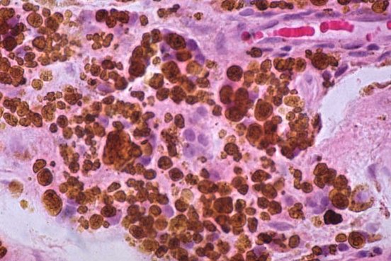
On
the left is an electron micrograph of a mast cell,
an
innate immune cell that mediates allergic responses.
On
the right, you can see the granules inside the mast
cell
that contain histamine, bradykinin, and other mediators.
When
IgE and an antigen crosslink on the surface of the
cell,
the granules release their contents into the
extracellular
space.
|
The reaction might remain local, but if it triggers the
same reaction throughout your circulatory system, it can cause anaphylactic shock, a true medical
emergency characterized by low blood pressure and respiratory difficulty. It
can and will kill you if not treated immediately. And all this because some
innocuous small molecule and an IgE antibody caused your immune system to over
react!
Type II
hypersensitivity reactions are also mediated by antibodies (IgM or IgG
type). The triggering antigen might be some foreign molecule bound to a host
cell or even an antigen on your own cells that your body has mistaken for
foreign. In the case of penicillin allergy, the drug becomes bound to your
cells; this complex triggers the immune response. If the antibodies are
directed toward your cells or mistake your cells as foreign, this is called an autoimmune reaction. Examples could be
systemic lupus erythematosus (SLE), some type I diabetes, or Hashimoto’s thyroiditis.
In some type II reactions, the antibodies that bind to the
antigens trigger the complement system in your tissues to activate. Complement
is part of your innate immune system that ends up marking cells for destruction
by phagocytosis, or destroys them itself by punching holes in the target cells. In some
cases, the antibodies bound to the cell trigger innate immune cells called natural killer lymphocytes – you can
guess what they do to the target cell. I guess everyone is a natural born
killer on the inside.
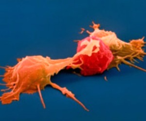 |
Natural
killer cells are lymphocytes, but are part
of
the innate immune system. These two are
attacking
a cancer cell (red). Natural killers
specialize
in killing cancer cells and virus-infected
cells.
Natural killers are unique in that they can
recognize
stressed cells in the absence of binding
antibodies.
|
Type IV
hypersensitivity is the exception; this response can take several hours to
develop and is the only hypersensitivity reaction that does not involve
antibodies. Lymphocytes of the adaptive immune system interact with the antigen
(be it foreign or domestic) and release many chemical mediators, called cytokines, that mediate immune and
inflammatory reactions. Allergic contact dermatitis from poison ivy is a
common, but relatively benign, example of this type of hypersensitivity.
Most hypersensitivities are reactions to things that
shouldn’t have been problems in the first place. Allergies are just the most
common manifestation of immune hypersensitivity. I don’t have them to any
degree, but I see the havoc they wreak on my wife and our son. He is so allergic
to wool that he breaks out when he counts sheep in bed!
But even allergies might have a hidden benefit. A study in 2008 indicated that
people with allergies actually have a 25% less chance of developing a certain
type of immune cell cancer, called B-cell non-Hodgkin’s lymphoma (NHL). If that person
has three different allergies, they are 40% less likely to develop NHL.
This seems amazing, but it is supported by a 2011 study showing that
people with allergies are 25% less likely to develop a type of brain tumor
called a glioma. Glial cells protect and support the neurons in the brain;
abnormal growth of these cells can lead to pressure and death of brain cells. Still think allergies are annoying?
 |
Sneezes
leave your mouth at over 100 miles and
hour
and can spread droplets over 30 feet. Sneezes
may
help get ride of unwanted antigens, but other
people
don’t want them either, so cover your mouth.
Sneezing
into the crook of your elbow is best for
limiting
spray and contamination – I saw it on
MythBusters.
|
Learning that allergies might prevent cancer may make you
less likely to take that antihistamine capsule. In fact, the treatment for all
immune hypersensitivity reactions involve avoiding the molecule,
removing the offending antigen and antibodies, and/or suppressing the immune
system. We take corticosteroids, antihistamines, and other drugs to prevent the
actions that might be saving us from cancer. However, you can help protect yourself without
drugs as well—just catch a parasitic infection.
Parasitic worm infections, whip worm (Trichuris trichiura) or
schistosoma for example, have a tendency to dampen the immune response, and can
prevent some relapses in autoimmune diseases such as multiple sclerosis. A 2005 study indicates that some success has been
had after dosing Crohn’s disease patient’s with intestinal worms.
 |
Meet
Pediculus humanus capitis, the common
head
louse
magnified only 80x. It is an ectoparasite,
meaning
it lives on the host, not in the host. They
have
been around for a long time; they have been
found
on Egyptian mummies. This is why most
Egyptians
shaved their heads and wore wigs.
|
Parasites seem to have evolved specific mechanisms that inhibit the reactions that would eliminate them from the host, so they dampen
immune responses as a defense. The mechanisms have not been worked out and may
be parasite specific. Even malarial and leishmaniasis parasites can suppress
the immune response, but I don’t recommend that you contract a deadly infection
just to alleviate your allergies.
These last studies suggest that we may be living too cleanly
– let’s take a look at that next week.
Calboli FC, Cox DG, Buring JE, Gaziano JM, Ma J, Stampfer M, Willett WC, Tworoger SS, Hunter DJ, Camargo CA Jr, Michaud DS. (2011). Prediagnostic plasma IgE levels and risk of adult glioma in four prospective cohort studies. J Natl Cancer Inst. DOI: 10.1093/jnci/djr361
Joseph A Jackson, Ida M Friberg, Luke Bolch, Ann Lowe, Catriona Ralli, Philip D Harris, Jerzy M Behnke, Janette E Bradley (2009). Immunomodulatory parasites and toll-like receptor-mediated tumour necrosis factor alpha responsiveness in wild mammals BMC Biology DOI: 10.1186/1741-7007-7-16
For more information or classroom activities, see:
For more information or classroom activities, see:
Allergy
–
Immune
hypersensitivity –
Autoimmune
diseases –