Wednesday, June 25, 2014

They Can See The Blood Running Through You

Biology concepts- thermosensors, TRPV1, hematophagy, taste sense, alternate splicing, echolocation


All three species of vampire bat live in Central to South
America, the common vampire bat (Desmodus rotundus),
the hairy-legged vampire bat (Diphylla ecaudata), and
the white-winged vampire bat (Diaemus youngi).
Any idea what the picture to the left shows? A hint – this may be the most sophisticated piece of machinery ever devised by nature. Together with the organism to which it’s attached, this piece of evolutionary engineering is capable of almost everything a billion dollar jet can do.

It’s the nose of the common vampire bat (Desmodus rotundus). These bats belong to the family Phyllostomidae, one of three families of leaf-nosed bats (Rhinolophidae and Megadermatidae being the other two families). One of the exceptional skills mediated by this nose makes use of the same receptor that makes our mouths burn when we eat chili peppers. Vampire bats can detect the hot blood in your veins from far away!

It’s the noseleaves of the vampire bat that are so amazing, but maybe we should include the rest of the bat head as well. The ears, teeth, mouth, and eyes all work with the nose to give this bat some jet fighter skills.

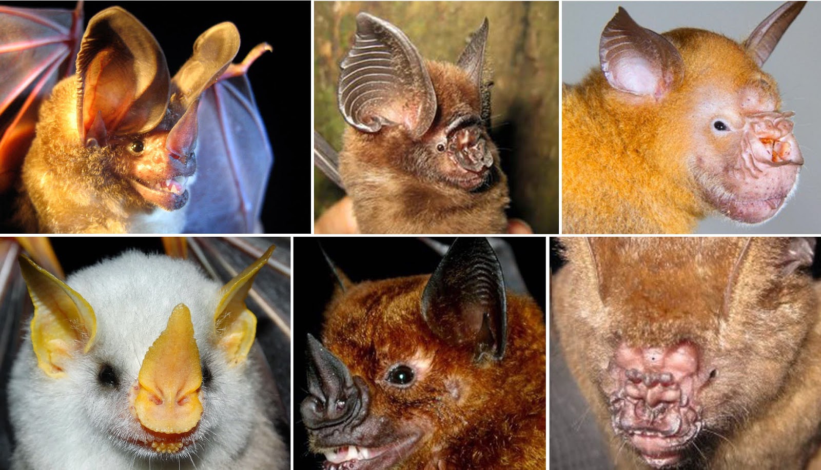
Leaf nosed bats come in some very odd varieties. The picture on the below and right will give you some idea of the shapes and sizes possible. The question is – what’s the reason for these bizarre growths and why isn’t one odd shape enough? The answer will best be found if we know what their function is, because in biology – form follows function (except for proteins, see this post).

Here are some different leaf nosed bats. Top middle is the Ridley’s leaf
nose bat; bottom left is the Honduran white bat. Top right is the
Commerson’s leaf nose bat and the middle bottom is the greater
spear-nosed bat. The bottom right image is of the cleaf nosed bat of
Vietnam, a more newly discovered variety. It was first described in
2008, but it took 4 years to determine if it was a new species or just
a variant of another species.

Two basic needs of the bat are to find food and find its way. Whether it's a fruit bat, an insectivorous bat, or a vampire bat, a bat must be able to negotiate obstacles within its environment and find a source of nutrition.

To accomplish these tasks, especially given that most bats are nocturnal, they use echolocation. They send out a high-pitched sound, and it bounces off objects and returns to their ears. This is very much like the radar used in airplanes. But this isn’t all they use. Bats can see just about as well as humans; the phrase “blind as a bat” might as well be “blind as a Bob.”


We said most bats are nocturnal. This is Livingstone’s fruit bat
or Comoro flying fox. It is a fruit eating bat of the Comoros
Islands in the western Indian Ocean (just northwest of Madagascar)
and is at least partially diurnal. Other bats may be seen during
the day, but it almost always because they have been disturbed
in their hiding place or they were disturbed in their
feeding the night before.
Bats can also smell their way to food, especially fruit bats. But to answer the question about the noseleaves we have to return to echolocation. Vampire bat sounds emitted for echolocation come through their nose, not their mouth! According to a 2010 study, the leaves aren’t used to gather the returning sound, but to focus the outgoing sound so that the “pictures” formed by the returning echos will be most accurate.

Different shapes help to increase the difference in the reflectivity of objects in the area of focus as opposed to those in the periphery. This allows the various species to hone in what they need to discern and dismiss those things that are uninteresting. Different backgrounds and different needs require different nose leaf shapes.

This answers the question about the wild shapes of noses, but it brings up another question. If vampire bats find their food by echolocation, sight, and smell, then why do they have heat sensors?

To answer this new question, consider the sizes of the vampire bat and its intended prey. The bat weighs about 2.5 oz (71 g), but it needs blood for food (mammalian blood for common vampire bats, bird blood for hairy legged and white-winged species). In fact, vampire bats are the only mammals that completely depend on hematophagy (blood meals). Because of this, they often feed on animals that are over 1000x their size.


Pigs are a favorite source of blood for vampire bats. Here, a
sleeping pig has been bitten on the snout. Why the snout – read
on. Notice the bat can hold its weight with its wings, and that
there seems to be more blood than you would expect from
such a small bite. Again, read on to know why.
To get to the blood of say a cow or horse, vampire bats would have to have teeth so large that they couldn’t lift themselves for flight. No, they have to be very careful about where they bite a victim; somewhere that will bring enough blood to feed on, but won’t cause the huge animal to kill them. Vampire bat teeth are so sharp that prey animals rarely feel the bite, and since the bats are nocturnal, the victims are most often asleep at the time and stay asleep during the feed.

The bats need to locate a place on the sleeping animals where blood vessels are near the surface. This is where the heat sensing comes into play. Vessels close to the surface will give off the most heat to the environment, and vampire bats can “see” these vessels from up to 20 cm away!

The vessels in question need to be covered with less hair, so the bat almost always goes for the lower leg or snout. They will land on the ground, and walk or run up to the prey from behind the animal to make the bite. Vampire bats wings are much stronger than most other bats, so they have an easier time moving along the ground, supporting some of their weight on their wings.

On the left you can see the incisors of the vampire bat. The cheek
teeth and canines are used to shave off any hair from the site, but
the incisors do the cutting. The lack enamel, so they are always
razor sharp. On the right, the tongue is being used to take in the
blood. The tongue is deeply grooved, so the anticoagulant saliva
runs down into the wound and more blood can easily be lapped up.
Their teeth then cut a 5 mm x 5 mm gouge in the victim and they lap up the blood that comes out. It isn’t too common, but vampire bats do feed on humans. This leads us to another amazing skill.

Instinct tells the vampire that a good feeding once will probably mean a good feeding again – if they can find the same animal. So how do they find the same animal several night is a row? They hear them.

A 2006 study showed that vampire bats do tend to feed on the same individual (be it human or cow) for several nights in a row. They can distinguish their previous victim by the sounds of their breathing! Every animal has a unique breathing pattern and sound profile, and the vampire bat can distinguish between individuals to find the one that matched a previous good meal. Imagine if we could find our favorite meal again by listening for the clinking of the right pans!

Returning to a good feeding spot each night, the vampire bat searches for a surface vessel to drink about 1-2 teaspoons of blood (4-5 ml). This isn’t enough to harm the animals, and is what allows them to go back several nights in a row.


Rabies can be spread by bats, and they don’t have to bite you.
When a bat bites an infected animal, it takes in the virus. The
virus grows in the animal and gets distributed to the saliva as
well. Startled bats sometimes spit, and if this gets into your
eyes, mouth, nose, or an open wound, you could contract the
infection. It’s rare overall, but rabies kills about 60,000
people a year.
This doesn’t mean that feeding by vampire bats is without negative consequences. One, the idea of being fed on gives me the heebie jeebies. Two, the vampire bat is a common vector for rabies virus. And three, in the cattle of Latin America, repeated feeding by vampire bats is associated with reduced milk production in dairy cattle and reduced mass gain in beef cattle. So if you wake to find a vampire bat licking your ankle, best to shoo him away and try to breathe differently tomorrow night.

How do vampire bats locate that ankle vessel they need to feed on? Back we go to that amazing nose. The heat sensors of bats are called pit organs, just like in the pit vipers we talked about last week. There are three to four of these organs in the noseleaves of the bat, and a couple across the upper lip as well.

As opposed to the pit vipers, vampire bats have adapted a heat sensor, not a cold sensor to use as their infrared detector for blood vessels. TRPV1, the same receptor that is used for the capsaicin burn and heat regulation in mammals, is present in very high numbers in the neurons of the pit organs.

But this is no ordinary TRPV1. Mammals can’t detect heat from 20 cm away with a regular TRPV1 – this is a modified TRPV1.  A 2011 study found that this version of the protein is missing the last three amino acids on the carboxy terminus (the end produced last). This small change increases the sensitivity of the receptor from 43˚C all the way down to 30˚C, so that small differences in heat can be noted from almost a foot away.

One more amazing fact - the bats have regular TRPV1 too. The two version of the protein come from the same gene and the normal one is used throughout the bat’s body for all the things we use TRPV1 for: heat regulation, reproduction, cancer inhibition, etc. Only in the neurons of the pit organs is the mRNA altered after it is transcribed from the gene (alternately spliced) to make the slightly shorter, more sensitive protein.


Here is a cartoon of how blood clots. On the bottom flow chart,
the first anti-co line is where desmolaris and draculin work.
The third line is where desmoteplase acts.
Now our bat friend has located a victim, found a surface vessel and taken a bite to let the blood flow. There’s yet another problem. Mammalian blood clots to prevent loss. The bats must either keep biting, which might wake their prey, or have a way to keep the blood flowing.

Their mouths have specialized salivary glands that make anticoagulants so no clot is formed. There is one anticoagulant that someone with a sense of humor named draculin. It acts to prevent blood clot formation. We have mentioned a second anticoagulant before, called desmoteplase. One of our Halloween posts talked about how it may be good for people that have had strokes. It dissolves any clots that may form.

A 2014 clinical trial is showing that desmoteplase is better than the tissue plasminogen activator clot busters now being used (rtPA), since they have a half-life of four hours (as opposed to 5 minutes for rtPA) and it’s breakdown products aren’t as toxic to nerves and the blood brain barrier as compared to rtPA.

A newer anticoagulant is called desmolaris. A 2013 study showed that it works on yet another part of the clotting system to prevent clot formation. And this isn’t all of them. A 2014 protein survey suggests that there may be dozens more anticoagulant proteins in vampire bat saliva.
Which flying machine is more complex and cool?

Lets add up the vampire bat’s technologies and compare them to an F16. The bat can fly and turn better. The bat has radar and infrared heat detection. It has high powered listening devices that can discriminate between two individuals. Finally, it has biological weapons that allow it to do its work without alarming the target.

All that in a “machine” that can fit into the palm of your hand. Defense aeronautical engineers must feel so embarrassed.

Next week, let’s take it just a bit further. Female mosquitoes aren’t just looking for you, they’re tasting and feeling for you. They use CO2 gradients as well as my prodigious heat to find me on a warm picnicking evening.


Vanderelst D, De Mey F, Peremans H, Geipel I, Kalko E, & Firzlaff U (2010). What noseleaves do for FM bats depends on their degree of sensorial specialization. PloS one, 5 (8) PMID: 20808438

Patel R, Ispoglou S, & Apostolakis S (2014). Desmoteplase as a potential treatment for cerebral ischaemia. Expert opinion on investigational drugs, 23 (6), 865-73 PMID: 24766516

Ma D, Mizurini DM, Assumpção TC, Li Y, Qi Y, Kotsyfakis M, Ribeiro JM, Monteiro RQ, & Francischetti IM (2013). Desmolaris, a novel factor XIa anticoagulant from the salivary gland of the vampire bat (Desmodus rotundus) inhibits inflammation and thrombosis in vivo. Blood, 122 (25), 4094-106 PMID: 24159172

Gröger U, & Wiegrebe L (2006). Classification of human breathing sounds by the common vampire bat, Desmodus rotundus. BMC biology, 4 PMID: 16780579

Gracheva EO, Cordero-Morales JF, González-Carcacía JA, Ingolia NT, Manno C, Aranguren CI, Weissman JS, & Julius D (2011). Ganglion-specific splicing of TRPV1 underlies infrared sensation in vampire bats. Nature, 476 (7358), 88-91 PMID: 21814281


For more information or classroom activities, see:

Leaf-nosed bats –

Echolocation –

Alternate splicing –

Anticoagulants -

Wednesday, June 18, 2014

Sneaking Up On A Snake

Biology concepts – thermosensor, sight-hunters, snake hearing, mutation, TRPA1, pit vipers

We have been talking about taste sense for many weeks. I
remember a 1975 movie called, A Boy And His Dog, starring a
very young Don Johnson. It was a post-apocalyptic story of a
guy, his dog, and cannibalism. The best line of the movie? “Well,
she might not have had good taste, but she sure tasted good.”
Of course, this isn’t the kind of tastes we have been talking about.
We’ve come a long way since we started talking about taste sense. We have learned about how TRPV1 capsaicin receptors sense pain and heat. We have also learned that TRPV1 capsaicin receptors have cousins that sense cold - TRPM8 and TRPA1. They may generate pain, and they certainly help to warm us when we are cold.

We have even learned that in rare cases, the cold receptors can be heat sensors, like in chickens and insects where TRPA1 sense hot instead of cold. And this leads us to today’s exception. It’s time to talk about how these relatives of taste receptors help animals to become better hunters and to better sense their environment. Today let’s focus on snakes.

Snakes have a number of ways to catch prey (see this post). Some lie in wait, blending in with the jungle or background until a moving potential dinner catches their eye and moves across their path. Vision is their primary way of finding dinner. As a consequence, most sight-hunting snakes are diurnal (active in daylight).

Here is the southern black racer. You can see it has big eyes with
round pupils so lots of light can enter – it’s a sight hunter. Many
grow to be 5 ft. (1.5 m) long, so they can look intimidating. But
they are not venomous and will usually exit the seen if disturbed.
The non-venomous Southern black racer (Coluber constrictor priapus) is a sight-hunting snake of North and Central America. It’s called a racer because it is quick, reaching 4 mph (1.8 kph) in a very short time. Even though it is a constrictor, it typically doesn’t coil around the lizard, mole, or bird (I said they were quick) that it catches. It prefers to crush them into the ground to suffocate them. Sometimes nature can be a little rough around the edges.

Other snakes use the combination of scent and taste that we talked about a while back. The Jacobson organ (more scientifically called the vomeronasal organ, VNO) in their mouth can sense the molecules that the tongue pulls in from the air. Like it or not, every organism has molecules floating off of them continuously. Snakes' VNO can pick these up. See this post for more on the VNO.

Some snakes “hear” their prey coming. True, snakes don’t have an outer ear opening or the small bones that convert sound waves into mechanical waves in our middle ear (see this post for an explanation). But they do have a cochlea, the organ for sensing the vibrations and converting them to a nerve signal. Many snakes can sense the vibrations that their prey generate when they move through the environment using this cochlea and their lower jaw.

Similar to something called bone conduction hearing in animals with ears like ours, vibrations that travel through the bone can also cause movement in the hairs of the cochlea. As we discussed previously, the bending of the sensory hairs of the cochlea are transduced to chemico-electrical signals that travel to the hearing centers of the brain.

This is from a scientific paper showing the bone hearing of a python.
The red is the lower jawbone. The bark blue is the quadrate bone
and the green is the equivalent to our stapes bone of the middle
ear. The light blue is the inner ear space and the purple is where
the cochlea is housed. Vibrations go from red, to blue, to green to
light blue, to purple. You can see how sound waves would find it
tough to get to the cochlea.
A 2008 study showed that many snakes rest their jaw bones against the ground. The vibrations caused by moving animals are transferred from the ground to the bone, and from the bone to the buried cochlea. The sensation in the brain is a lot like muffled knocks, not unlike the bass that is turned up too loud in peoples’ cars.

This was followed by a 2012 study that showed pythons have very sensitive vibratory hearing, but poor sound pressure hearing. Almost all their hearing input comes from the vibrations they sense in the ground or tree, or whatever they happen to be lying on. So be on tip toes, that snake may hear you coming.

But how does any of this relate to a receptor for painful cold and controls mammalian breathing rate? Well, another way some snakes find their prey is by sensing the heat they give off – even from a few meters away.

Pit vipers are a subfamily of the Viperdae family, called Crotalinae. There are two types of vipers; all of them have hinged fangs, the ones that are folded up into the upper jaw when the mouth is closed, but protrude for striking as the mouth is opened. Pit vipers differ from true vipers in that they have pits (duh!); more about these below. True vipers live exclusively in Africa and tropical Europe and Asia.

In America, where I live, there are a lot of pit vipers. Cottonmouths, rattlesnakes (all 30 species), water moccasins, copperheads – these are all pit vipers. From southern Canada to Argentina, and from Eastern Europe to parts of Asia, pit vipers are not rare. Eyelash vipers (Bothriechis schlegelii) of South America are arboreal (live in the trees). They have bright coloring, but sit still and wait for their prey to happen by. They strike from above, so they scare the heck out of jungle hikers.

On the left is the eyelash viper. You can see it doesn’t mean business
because its hinged fangs aren’t extended. In the middle is the two-striped
forest pit viper. It is protecting it’s young, so the fangs are extended. On
the right is a sidewinder rattlesnake. Sidewinders are amazing and will
get their own post soon.
The amazing thing is that there aren’t any pit vipers or true vipers in Australia. The land of a million weird and painful deaths has nothing to offer in the way of hinged fang venomous snakes. I’m sure there’s a movement to import some.

But it’s specific part of the pit viper that we are interested in today – namely the pit. The pit organ is located between the eye and the nostril, on each side of the snake’s head. It is a hollow pit, so the actual business end of the pit organ is inside the snake’s skull.

The pit is lined with epithelium, but it also has a membrane that is stretched across the base. As a consequence of the location membrane, there are air pockets on each side of the membrane. The trigeminal nerve innervates the membrane and there are thermosensors in the cells of the membrane.

So, the pit organ is a thermosensor that helps them locate prey animals (or predators). But wait you say. Sure, pit vipers may use a thermosensitive ion channel to sense the heat given off by passing prey animals. But we just said they use a COLD sensing ion channel, TRPA1. What gives?

The pit on a pit viper is located between the nostril and the eye.
It would be easy to mistake the pit for the nostril. The cartoon
shows the pit anatomy. The air chamber helps cool the air
quickly and stops the TRPA1 receptors from firing again. This
is so the snake won’t get a residual image of something warm,
when the target may have moved in the interim period.
The explanation is two fold. 1) We said a couple of weeks ago that TRPA1 might sense painful cold on its own, or may work with other TRP’s to respond to very cold temperature. But whichever way it works, it is very similar to TRPV1 for heat sensing and TRPM8 for cold sensing. 2) Remember that in birds, lizards, and many insects, TRPA1 actually senses heat, not cold.

So maybe it’s not so terribly bizarre that pit vipers use TRPA1 to sense their prey. But before they touch it??? We eat chili peppers and we react to the capsaicin in our mouths and noses. We go out on a summer day, and the heat activates our TRPV receptors in skin and other tissues. We eat something cold (or menthol) and we feel the cold sensations it touches or tissues. But snakes feel the heat of their prey before they eat, from a distance away! There must be more at work.

And there is. The TRPA1 ion channels in the pit organs of pit vipers have a mutated version of TRPA1. Here’s how things work according to a 2010 study that identified TRPA1 as the heat sensor. The pit is a hole with a membrane stretched toward the back. Consequently, there is an air chamber on both sides of the membrane.  The membrane is highly vasculature and has the sensitive nerve endings with the TRPA1 channels.

The TRPA1 receptors are always firing, but at a low rate. Neutrally warm objects don’t change the firing rate, but warmer objects (as little as 0.001 ˚C warmer than background) will increase the firing rate. The receptor is mutated according to a 2011 study, with 11 amino acids of the pit TRPA1 divergent on only pit-containing snakes. These changes make the receptor so sensitive that it can react to infrared light signals (heat) from several feet away. That would be like our mouth burning over a chili pepper that we walked past in the supermarket.

Since the sensors are spread across the entire membrane, the effect on locating the source is sort of like vision or a pinhole camera. Light passes through the pupil and diverges before it hits the retina. This provides for a larger spread of the “image” across the membrane and allows for precise two-dimensional map of the target. The difference in heat between the target and the background gives a “picture” of the object that is warm.
The Taylor’s Cantil viper will play dead and then strike, but this
brings up an important point. DON’T get near a pit viper, even if
you are sure it’s dead. The pit is wired directly to the brain and
muscles. A dead snake, even one with a severed head, can still
strike as long as there is any residual neural electrical flow. People
die every year from snake bites from dead snakes.

The picture generated is also a little like hearing, since the heat will reach one pit earlier or more strongly. By comparing the timing and the strength of the signals from each pit, the distance and direction to the target can be detected by the brain (see this post for localization of sound waves).

Because the heat “picture” pit vipers pick up is based on the difference between the temperature of target and background, most pit vipers hunt when coolest, so temperature gradient between environment and prey is greatest. Prey will stick out the most.

Snakes can also use the pit more conventionally, as a thermosensor for its whole body. The basal rate of firing will tell the snake when to move to shade if it’s too warm or move to sun if it’s too cold. This is how it regulates its body temperature.

Pythons and boas can also have heat-sensing pits, but they are
5-10 times less sensitive because of their differing anatomy.
The amazing thing is that they evolved the same special power
independently from pit vipers, although they both use mutated
versions of TRPA1. The nostril has a black arrow and the pits
have red arrows.
The exception to today’s exception: some non-pit vipers have pits. In terms of evolution, pits evolved once in pit vipers, but they have sprung up several times in boas and pythons. These pits are less sophisticated (no membrane or air chambers), are less sensitive, and are located in different places.

Boas and pythons with pits have 3-4 simple pits in their upper lips. They don’t have the suspended membrane for sensing temperature, the TRPA1 sensors are housed within the epidermal cells at the back of the pit.

Next week – vampire bats and mosquitoes get into the mutated thermosensor act as well.


Christensen CB, Christensen-Dalsgaard J, Brandt C, & Madsen PT (2012). Hearing with an atympanic ear: good vibration and poor sound-pressure detection in the royal python, Python regius. The Journal of experimental biology, 215 (Pt 2), 331-42 PMID: 22189777

Gracheva EO, Ingolia NT, Kelly YM, Cordero-Morales JF, Hollopeter G, Chesler AT, Sánchez EE, Perez JC, Weissman JS, & Julius D (2010). Molecular basis of infrared detection by snakes. Nature, 464 (7291), 1006-11 PMID: 20228791

Geng J, Liang D, Jiang K, & Zhang P (2011). Molecular evolution of the infrared sensory gene TRPA1 in snakes and implications for functional studies. PloS one, 6 (12) PMID: 22163322


For more information or classroom activities, see:
Pit vipers –

Bone conduction hearing –

VNO (Jacobson organ) -

Wednesday, June 11, 2014

What Cold Really Looks Like

Biology concepts – TRPA1, cold sensing, oxygen sensing, proteasome function, hypoxia, normoxia, hyperoxia, phototransduction, optogenetics, methyl anthranilate

Last week we learned that the TRPA1 ion channel causes you pain when you are too cold and helps you to avoid the cell damage that cold can produce. Even if that’s all it did, it would be pretty amazing, but there’s much more. It turns out that TRPA1 can do some amazing things - like keeping the right amount of oxygen in your cells.

Prolyl hydroxylase domain enzymes (PHD1, 2, and 3) are the primary mammalian sensors for oxygen in the blood and tissues when oxygen levels are normal (normoxia) or low (hypoxia). Oxygen binding to the PHD acts as an "on" switch for the binding of another molecule to PHD, a molecule called 2-oxoglutarate (2-OG). 2-OG can only bind to PHD if O2 is already bound.


On the left is a cartoon that shows how ubiquitin and PHD proteins
help mediate the destruction of unneeded damaged proteins. On the
right is the proteasome, the structure which recognizes the proteins
targeted for destruction and carries out the cutting that
releases amino acids.
The function of 2-OG bound to a PHD is to trigger the destruction of another type of protein, called hypoxia inducible factors (HIFs). They do this by modifying some of the amino acids of the HIF so that cell thinks the HIF is old or defective. Old and defective proteins are recycled for their parts by a big complex of proteins called a proteasome.

When there is sufficient oxygen in the blood and tissues, the PHD is bound by O2 and 2-OG, so the HIFs are degraded. But when there is hypoxia – little O2 and 2-OG are bound to the PHD – and there is no destruction of HIFs.

If they aren’t targeted for the proteasome, HIFs are free to do their job. They turn on genes that work to increase oxygen in the blood and tissues, including making more red blood cells, using more iron for heme, stimulating the production of new blood vessels, and triggering cells to use glycolysis instead of the citric acid cycle because glycolysis doesn’t need O2 (here’s a review).

When oxygen levels in blood and tissue return to normal, more O2 will be free to bind to the PHDs and the HIFS will then be degraded. Their effects on genes will be turned off as their concentration decreases.


Oxygen sensing in the cells is important at all stages of life. At one
time it was believed that very premature infants, with less than
mature lungs, would benefit from a high oxygen environment.
What it actually did was make them blind due to oxygen mediated
damaged to the retina.
This is all well and good for normoxic or hypoxic situations, but how would it help in times of too much oxygen (hyperoxia)? And believe me, too much oxygen is a very bad thing. Short bursts of high oxygen can be disorientating, with central nervous symptoms that affect breathing and may cause myopia.

Prolonged exposure to high levels of O2 (or short exposures to very high levels) can cause cell destruction, collapse of lung alveoli, retinal detachment, and seizures. Scuba divers, firemen, and anyone else using oxygen tanks must be aware of the dangers of either running out of oxygen or breathing in too much.

This is where TRPA1 ion channels enter the picture. A 2008 study showed that TRPA1 can be activated by O2. In hyperoxic situations, there is too much oxygen to be bound up by the available PHDs, so some is left to interact with TRPA1 channels in the membranes of the vagus and sensory neurons, as well as in tissue cells. The O2 can open the TRPA1 channel directly and lead to firing of the neuron.

The more O2 there is, there more activation of TRPA1, and this is good. In hyperoxic situations, the body constricts many blood vessels to limit the excess oxygen getting into the tissues. Hyperoxia also ramps up the cells’ protective mechanisms against reactive oxygen species damage.

 What we don’t know is which responses to hyperoxia are controlled by the TRPA1 channel activity.  A 2011 study goes onto show that the PHDs lose their inhibitory function on TRPA1 in both hyperoxia and hypoxia. This is a similar conundrum to the one we saw last week - where TRPA1 senses cold, but responds to many of the “hot” agonists of TRPV1. Here, the same sensor is stimulating different responses to too much or too little oxygen.


Some situations with low oxygen tension can create big problems.
On the left is pneumonia; the filling of the alveoli with edema fluid
limits the amount of oxygen that can get to the bloodstream. In the
middle is Payne Stewart; at altitude, his plane lost cabin pressure,
everyone on board basically went to sleep, and the plane flew in a
straight line until it ran out of fuel and crashed. On the right is a diver
who specializes in going as deep as possible on one breath. Many
participants drown.
The same channel that senses really cold temperatures in your skin and tells you that this bad by stimulating pain also helps you keep a normal amount of oxygen in your tissues – a system that has little or nothing to do with pain. Weird enough for you? Well it gets weirder.

It seems that your TRPA1 cold sensing channels are important for tanning in the summer! A 2013 study shows that a process called phototransduction uses G-protein coupled receptors to stimulate TRPA1 in melanocyte and keratinocyte membranes and results in an influx of calcium. This destabilizes the membranes and facilitates the transfer of melanosomes from melanocytes into the surrounding keratinocytes, as we have talked about before.

Phototransduction in general is where light energy is changed (transduced) into another form of energy. The best example is in the retina of the eye, where rods and cones turn visual light into neural signals that are then processed in the brain as images – that’s how you see. In the skin, the energy of the UV light is turned into chemical energy (flow of ions in and out of channels) to stimulate cellular activity.


The retina is the most obvious example of phototransduction. Light
energy is converted to chemical energy and information. Light
strikes the retina, and excites the rods and cones. On the level of
the membrane, the light (hv) set in motion a series of reactions that
results in the opening or closing of ion channels, including
the TRPA1 channel.
A second 2013 study from the same group says that retinol (hear the word retina in there? As in the retina of your eye?) is the photoactive chemical that starts the G-protein couple receptor cascade that then results in TRPA1 activation and melanin synthesis in melanocytes.

A new photosensitive protein called optovin has been identified in zebrafish. It mediates TRPA1 activation via a sensitive part of the TRPA1. Optovin allows for optical control of TRPA1-expressing neurons, meaning - optovin absorbs light, generates singlet oxygen radicals and these interact with the the oxygen-sensitive cysteine residues on TRPA1 and activates the receptor. Sound familiar? This is similar to the binding of oxygen that helps the body recognize and respond to hyperoxia.

In an effort to take advantage of this great system, the research field of optogenetics has been born. Let’s say you want to study what happens when a set of neurons fires. Introduce optovin and TRPA1 (or a similar system, like opsin or retinal) into the appropriate neural pathway and then you can fire them at will just by shining a light on them. Imagine, shine a laser pointer on a mouse and instantly he starts to jump, or drool, or tell a joke.

Optogenetics was the method of the year in 2010! On the left
is how it works, using phototransduction molecules to stimulate
responses in target cells. On the left is how it works in a live rat.
The light is positioned so that it can illuminate the altered
neurons so that processes can be turned on and off with light.

One last exception for the day – one that will lead to some amazing stories for next week. In mammals and many other animals, TRPA1 senses noxious cold (in addition to the amazing things we just talked about), but in some species it acts completely the opposite.

In mosquitoes, TRPA1 senses heat instead of cold. A 2013 study shows that larval A. gambiae (the mosquitoes that carry malaria) rely on TRPA1. If you decrease the amount of TRPA1, the mosquito larvae don’t exhibit thermal locomotive behaviors that would normally keep them in the preferred temperature of water. This might be important for killing mosquito larvae in water; if you can use antagonists to TRPA1 to move them away from their optimum temperature, they’ll die as babies and never become bloodsuckers.

In fruit flies, TRPA1 is also important for circadian (daily) locomotor activity patterns (2013 study). Instead of having a light/dark drive their activity, the temperature fluctuations between day and night can also serve to entrain circadian cycles. There are activity cells in fly brain for morning, daytime, and evening activity levels. TRPA1 isn’t expressed in morning neurons, so it’s activation by heat is what increases activity in day and evening.


TRPA1 gene function (painless in fruit flies) controls many
circadian behaviors. Depending on the amount of firing, it
tells the fly what time of day it is, and this time controls
which behaviors will be favored.
Another 2013 study shows that TRPA1 is used as thermoregulatory control of circadian rhythm in drosophila. Loss of TRPA1 altered behaviors and changed the expression of an important circadian rhythm protein called Per in the pacemaker cells.

In this one regard, birds are a lot like mosquitoes and flies. Chickens for example, have TRPA1 channels that induce pain due to high heat, just like their TRPV1 channels (which, you remember, don’t react to capsaicin).

A 2014 study showed that chicken TRPA1 is a heat and noxious chemical sensor – it acts opposite to the TRPA1 in humans, even though we are both homeotherms (maintain a body temperature within a small range). Chicken TRPA1 is almost always co-expressed with TRPV1, so they double up on heat but might have less cold sensing – why? - cold can still be damaging to cells so they need to know to avoid it.

The weird TRPA1 of birds lends itself to a bizarre use for us. Methyl anthranilate (MA) is a non-lethal bird repellent, and the same 2014 study shows that it works by activating bird TRPA1 pain sensors. MA doesn’t activate TRPA1 in other species, like humans; three amino acids critical for its MA activity are different in bird and mammal TRPA1.

Since MA doesn’t work on human TRPA1, it can be sprayed on crops to keep birds away – it’s a chemical scarecrow - if it only had a brain! It can also be sprayed on surfaces to keep birds from congregating. MA works as a repellent by stimulating the trigeminal nerves via TRPA1 in the bird’s beak, eyes and throat. 


Grapples (pronounced grape – ple) are apples soaked in
methyl anthranilate (MA) so they taste like grapes. MA is
sensed as hot in birds by TRPA1 but does not activate
the human TRPA1. We taste it as grape flavor and smell.
For me – if you want to taste grapes, eat grapes!
Very similar to MA is dimethyl anthranilate (diMA). It also activates bird TRPA1 and can be used as a repellent, but we use it for flavoring grape Kool Aid. Too bad Jim Jones wasn’t leading a flock of chickens – they’d all still be alive.

Both MA and diMA are naturally occurring in concord grapes, strawberries, other fruits, and are especially important for flavor of apples. Maybe that’s why they make grapples – grape-flavored apples. Actually, grapples are apples soaked in MA, not a genetic hybrid. DiMA is also released from musk glands of foxes, and is produced in rotting flesh – does spoiling meat taste like apples or grapes to you?

Next week – odd changes in TRPA1 and TRPV1 turn animals into better hunters.


Bellono, N., & Oancea, E. (2013). UV light phototransduction depolarizes human melanocytes Channels, 7 (4) DOI: 10.4161/chan.25322

Bellono NW, Kammel LG, Zimmerman AL, & Oancea E (2013). UV light phototransduction activates transient receptor potential A1 ion channels in human melanocytes. Proceedings of the National Academy of Sciences of the United States of America, 110 (6), 2383-8 PMID: 23345429

Saito S, Banzawa N, Fukuta N, Saito CT, Takahashi K, Imagawa T, Ohta T, & Tominaga M (2014). Heat and noxious chemical sensor, chicken TRPA1, as a target of bird repellents and identification of its structural determinants by multispecies functional comparison. Molecular biology and evolution, 31 (3), 708-22 PMID: 24398321


For more information or classroom activities, see:

Oxygen sensing –

Proteasome –

Phototransduction –

Optogenetics –

Methyl anthranilate –