Showing posts with label infectious disease. Show all posts

Wednesday, April 27, 2016

Your Body Has A Photographic Memory

Biology Concepts – innate immunity, acquired immunity, memory response, influenza

Your body is exposed to tens of thousands of foreign molecules every day. Some can do you harm, some can’t. Your immune system sorts them by matching receptors on immune cells to molecules on the foreign objects.

Legos and biology are a good fit. They can be used to analogize the 
rearrangement T cell receptor genes or hypervariable regions 
of antibody genes, or they can be used to model the entire 
body. One scientist uses them to model building 
complex systems from repetitive units. And they’re fun.
Think of the receptors as Legos; your DNA provides for several different types of Lego blocks to be made, and your immune cells can rearrange the different types and put them together as a receptor, so there can be millions of different receptors. Each immune cell has just one type of Lego receptor, although it may have many copies of that one form. Each different Lego receptor will fit, key in lock style, with a specific foreign molecule.

The receptors exist on many types of cells, and antibodies sometimes function as receptors when attached to the surface of specialized immune cells. Even circulating antibodies (Ab) in the blood take the form of key and lock systems, whether as single Ab, dimers (2) or pentamer (5) complexes.

The immune system of higher animals can be described as several sets of pairs. Each member of a pair attacks a problem in a certain way, and has independent pathways, but each pair also has overlap and must work together in an overall response. We could spend weeks just on this system, but lets look at the major parts by describing each pair, from largest to smallest.

Innate immunity vs. adaptive immunity – the innate immune responses are fast but short. They don’t depend on your immune system recognizing the specific foreign molecule (antigen) with a specific receptor, but respond with the same types of reactions no matter what it is. Almost all plants and animals have some form of innate immune system.

Vertebrates take the immune system further. They have developed an adaptive immune system that does depend on your immune system recognizing the specific foreign invader. It then generates a tailored response to that one foreign organism or molecule. The faster, but more general, innate response helps the slower, but longer lasting and more specific, adaptive response to kick in.

These are cartoons of an antibody. The model on the left is a much 
more realistic image. The Fc portion is the same through most 
antibodies (c= constant), while the gene rearrangement takes place 
in the light chain and heavy chain variable regions. The 
different variable regions are the Lego blocks that can be put 
together differently to make the millions of different antigen 
binding sites.
Humoral immunity vs. cellular immunity – when an antigen is recognized by an adaptive immune cell (often through antigen presentation by the innate system), an early response is for the cell to divide and make more of itself. You don’t get sick from one bacterium infecting you; many infect you at once and then divide to become many more. You need many copies of that specific immune cell in order to battle the invading horde of bacteria.

The immune cells can generate an antibody response (humoral immunity) and/or trigger specific killing and directing cells to be produced (cellular immunity). The antibody (produced by B lymphocytes) is a protein that recognizes the specific antigen. The cellular immune response is mediated primarily by T lymphocytes.

However, B cell-produced antibodies are important for T cells to do their work, and antibodies also help the innate immune response to keep working after specific recognition has been made. In addition, the cellular immune response can control and ramp-up the humoral response. You see what I mean about each pair being separate but connected.

Effector T cells vs. regulatory T cells – There are pairs of T cells as well. I use the term “effector T” cells to lump CD8+ and CD4+ lymphocytes together (CD = cluster of differentiation markers on the cell surfaces). Effector T lymphocytes are either directly cytotoxic (CD8+, cyto = cell and toxic = damaging) or command (CD4+) the many adaptive responses. Effector cells are contrasted with regulatory cells, which include regulatory and suppressor T lymphocytes. The purpose of these cells is to stem the effector response so it doesn’t get out of hand; parts of the immune response are inflammation and non-specific cell killing – too much of that and you die too.

Memory Immune System – This last part of the immune response is not a member of a pair. When your innate immune system is activated, it ramps up, does its job, and hopefully is turned back off. The adaptive immune system responds to the antigen by producing more cells, antibodies and chemical signals (cytokines), and after the invader is vanquished you want this response to diminish as well. The innate system always starts over from zero, but the adaptive system remembers the infection you had.

The dendritc cell on the left is an innate immune cell that works 
to present the antigen to the adaptive immune cells (Th1, 
Th2, and B cells). The adaptive cells reproduce and make 
cytokines to stimulate other immune cells. They also generate 
some memory cells that recognize the same antigen, but stay 
around for a long time and can react strongly and quickly.
During the adaptive response, some of the produced immune cells become “memory cells,” they still recognize the antigen from the initial infection, but hang around in larger numbers; in many cases they circulate in your body for the rest of your life. If your body sees that specific antigen again, the memory response can be re-initaited very quickly and very aggressively. You might be infected again, but your memory response is so fast and effective that you never know it.

In a world without vaccines, you are infected, get the disease, recover (hopefully), and then have a memory immune system for that antigen. Vaccines take the initial infection and disease out of the equation; you get to develop a memory without having had the experience!

As we discussed last week with smallpox, vaccines present your immune system with the antigen in the form of a dead or weakened pathogen, or just the antigen molecule itself. Your body doesn’t know the difference, it develops an adaptive and memory response just as if it were the real infection.

In the majority of cases, you develop memory B and T lymphocytes when infected or vaccinated. However, there are exceptions. Most antigens cannot fully activate B cells to make antibody, they have to be helped along by antigen-activated T cells. But there are T cell-independent antigens that can fully activate B cells on their own. In these infections, you can develop a B cell memory without a T cell memory.

On the other hand, there are other infections that develop a full memory response, but it is not useful. Influenza is an example of this. Influenza has been around for thousands of years; some years we have severe epidemics or even world-wide pandemics. The 1918-1919 Spanish flu pandemic killed over 50 million people, many more than the contemporaneous WWI (16 million deaths).

Flu is difficult to vaccinate against because it keeps changing. Influenza virus has two antigens, called H (hemagglutinin) and N (neuraminidase). These are the molecules on the virus particle that your body mounts an immune response against.

The H molecule on the viral coat binds to sialic acid receptors on respiratory cells and allows the virus to enter. When the newly produced viruses bud off of the cell, they place H on the cell surface, but there are still host sialic acid receptors there as well. These receptors would bind up the H and prevent the new viral particles from attaching to and infecting other cells, so the N molecule cleaves the sialic acid receptors from the new viral particles.

Influenza virus can mutate by antigenic drift or antigenic
shift. The top line shows that by passing from person to
person, the antigens (and virulence) shift slightly. The lower
line shows that by passing through other animals and
recombining, the antigens can have small or big changes. When
shifted virus moves into humans, it’s a recipe for a pandemic.
The problem arises when the H and N antigens mutate.... and they do. Scientists have identified 16 different classes of H’s and 9 different N’s, and they can be paired up in many combinations. Small changes (antigenic drift) usually mean that memory might have a slight protective effect, and major epidemics do not occur. But major changes in H and N (antigenic shift) mean that previously infected people have no memory protection.

Different strains of influenza virus can infect the same animal (often pigs and ducks – thus avian flus and swine flus) and can mix their H’s and N’s. What emerges and might be transmitted to humans can be a virus with H’s and N’s similar to years past, or with new H’s or N’s. That is why a new vaccine must be produced each year, after scientists see which H’s and N’s the new virus has and how much they have drifted. Avian flu is H5N1, while swine flu is H1N1. However, antigenic drift means that each H1N1 will not be exactly like the previous H1N1 to emerge. The 1918 pandemic was caused by an antigenically shifted H1N1 sub-strain.

Like flu, other infections may not provide life-long memory. If the memory response is weak or the initial response was not strong, then memory may fade over time. This is why some vaccinations require boosters in later years. A fading of the memory response to influenza is also implicated in the need for yearly vaccinations.

Here's a great book that discusses both the biology
and sociology of influenza. There are great personal
stories as well as medical detective work. This
pandemic was a jolt that brought infectious
disease research into a new century. I highly
recommend it.
Now for the exception to the exception. Influenza changes each year, so memory does not help much, but a 2010 report from scientists in Hong Kong suggests that prior exposure to any seasonal influenza (either by infection or vaccination) might have been a contributing factor as to why the 2009 pandemic of antigenically shifted swine flu (H1N1) was much milder than expected.

The 2009 seasonal flu vaccine did not have any cross-reactivity with pandemic H1N1, so the scientists suggest that previous years seasonal influenzas did generate some memory response that was partially effective against 2009’s H1N1 swine flu. Cross-reactivity means that the H and N antigens were not identical to previous version; the Legos don’t fit together exactly, but they were similar enough to fit together and initiate a partial response. Once again, we see that getting sick may save your life down the line.

Next week will look at examples wherein having one disease can protect you from catching another.


Mathews, J., McBryde, E., McVernon, J., Pallaghy, P., & McCaw, J. (2010). Prior immunity helps to explain wave-like behaviour of pandemic influenza in 1918-9 BMC Infectious Diseases, 10 (1) DOI: 10.1186/1471-2334-10-128

Kash, J., Qi, L., Dugan, V., Jagger, B., Hrabal, R., Memoli, M., Morens, D., & Taubenberger, J. (2010). Prior infection with classical swine H1N1 influenza viruses is associated with protective immunity to the 2009 pandemic H1N1 virus Influenza and Other Respiratory Viruses, 4 (3), 121-127 DOI: 10.1111/j.1750-2659.2010.00132.x

Cowling, B., Ng, S., Ma, E., Cheng, C., Wai, W., Fang, V., Chan, K., Ip, D., Chiu, S., Peiris, J., & Leung, G. (2010). Protective Efficacy of Seasonal Influenza Vaccination against Seasonal and Pandemic Influenza Virus Infection during 2009 in Hong Kong Clinical Infectious Diseases, 51 (12), 1370-1379 DOI: 10.1086/657311


For more information or classroom activities, see:

innate immunity:

adaptive immunity:

memory immune response:

influenza virus:
http://www.xvivo.net/zirus-antivirotics-condensed/

Wednesday, January 27, 2016

An Infectious, Genetic Disease? Better Sleep On It.

Biology concepts – thermoregulation, sleep, genetic disease, infectious disease, central dogma of molecular biology, form follows function


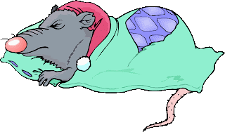
Even rats have to get some sleep. It was nice to have the sleeping cap,
but unnecessary for a sleep deprivation study. Not a good use of
research dollars.
“I’m dying for a good night’s sleep.” Is this just hyperbole, or an impending warning of death? For laboratory rats, sleep deprivation does kill. During their insomniac downward spiral, the rats tend to get hot and can’t cool down – you know, they can't thermoregulate (see Can’t We Just Go With The Flow). This doesn’t mean that a loss of the ability to thermoregulate is what kills the rats, but it does suggest a connection between sleep deprivation and the hypothalamus.

We looked at the hypothalamus in our story of endothermy. This evolutionarily old brain structure implements a set point temperature for the body and receives information about the temperature of different parts of the body. When the body temperature deviates from the set point, the hypothalamus initiates bodily mechanisms to normalize the temperature.


Apparently one of the effects of sleep deprivation is that you
become semi-transparent.
People with severe insomnia tend to sweat more and have higher core temperatures even though they say they are cold. They also have extreme high blood pressure, pulse, and appetite. These symptoms suggest that sleep deprivation messes with the hypothalamus, since functions of the hypothalamus include themoregulation, sleep, hunger, thirst, reproductive readiness in females, and stress responses. What scientists don’t know yet is just how sleep deprivation actually kills the rats or harms people.

Dying from a lack of sleep is not just a rat problem, a few very unlucky humans die from it as well. Fatal familial insomnia (FFI) is a very rare genetic disorder; it has been reported in only 40 families worldwide. Before describing the truly horrible way these patients die, let’s look at what causes the disease.

FFI is caused by a point mutation in the gene for the prion protein PrPc. A point mutation means that one nucleotide on the DNA is changed, which leads to a change in the protein coded for by the DNA. Three unit (nucleotides) segments of the RNA (made from the DNA template) work together (called a codon) to code for one protein building block (amino acid). In the case of FFI, the amino acid called aspartic acid is changed to one called asparagine, and this changes the protein’s shape. 


The left image shows mRNA bases recognized in sets of three
(codons) by tRNAs with amino acids attached (Ser = serine, tyr =
tyrosine). The amino acids are linked to because proteins. The
lower section is the genetic code, showing which amino acids are
coded for by which codons. The right image shows how proteins
fold. The primary structure is the amino acid sequence. The
secondary structure comes from interactions of adjacent amino acids,
including spirals called helices or sheets. The tertiary structure comes
from the folding up of the entire protein, while the quaternary
structure comes from the interaction of different proteins into a
larger complex.
PrPc is made up of 250 amino acids linked together in a chain. Each different amino acid carries a different shape and charge and will interact with every other amino acid differently. The sequence of amino acids in a protein cause it to fold into a specific shape. It is the protein’s conformation (shape) that determines its function. This is the opposite of what we determined for evolved organism characteristics, where form follows function (see Do You Have To Be Ugly To Hear Well?). With proteins – function follows form!

Mutation of that single amino acid at position 178 (aspartic acid is negatively charged, while asparagine is positive) causes the folding, and therefore the function, of the protein to change. Aspartic acid is sometimes abbreviated "D", while asparagine is called "N"; therefore, the mutation is often indicated as D178N (D at position 178 is changed to N).

Many genetic mutations result in no change in amino acid, or a change that bring a large enough change the shape to cause a change in function. But when it does, good or bad things can happen. On one hand, the altered protein might confer an advantage to the organism, one that promotes survival in the environment or after an environmental change.This positive selection through reproductive advantage become the new normal – and this is evolution

On the other hand, the change in amino acid sequence, form, and function could be destructive. Disease might be the result, or perhaps a change in the organism that reduces reproductive success. One of these two results is what occurs with the FFI mutation of the prion protein.

When the mutated prion folds differently, it forgets its day job and moonlights as a sinister evil force. Every other prion protein it contacts, WHETHER MUTATED OR NOT, is coaxed into changing its shape. The new prions turn to the dark side, then change other prion proteins they contact, multiplying the effect. The poorly folded prion proteins will stick together, come out of solution, and form solids (plaques) where they settle out. In different prion protein diseases, this settling out occurs in different parts of the brain. In FFI, it is the hypothalamus.


In the top image, the PrPc on the left is properly folded. The green
represents alpha helices and the blue arrows represent beta-pleated
sheets. The right image shows the malfolded version of PrPsc. It is a
tighter structure, which partially explains why protein-degrading
enzymes don’t work on it. . The lower cartoon shows that the PrPsc
can force the PrPc to assume the improper form, and these then
aggregate into plaques.
The prion plaques are longer lived then the regular prion protein; normal cellular enzymes whose job it is to degrade proteins won’t work on prion plaques. And worse, if some of the malfolded protein is transferred to another animal, the recipient will develop plaques and disease as well. That makes this an infectious disease that isn’t caused by a bacteria, fungus, parasite, or virus. The prion is an infectious protein! What a terrible exception to the rules of infectious diseases.

We see here a protein that can replicate itself (not by building more of themselves, but by changing the form of normal proteins), and that makes it a repository of biologic information. This is an exception to the central dogma of molecular biology, which says that DNA is the sole information storing material.

FFI moves from person to person through heredity, but if a non-affected person comes into contact with some brain material from an FFI patient and that material entered their bloodstream, it can be transmitted this way as well. A prion protein disease called Kuru is famous for being transmitted from person to person.

The Fore tribe in Papua New Guinea once observed a ritual wherein they honored a dead tribe member by eating part of their brain (called ritualistic mortuary cannibalism - gasp!). Because of this, there was an epidemic of Kuru in this tribe in the early 1900’s. Over a period of 3-6 months victims would become unsteady, irrational with bouts of laughter, and then degrade mentally and physically to the point of death. There are more than twenty known prion diseases (mad cow disease, Creutzfeldt-Jakob, scrapie, etc.), and Kuru suggests that some might have no genetic component, only person to person transmission.


A member of the Fore tribe is shown on the left. This tribe used
to celebrate the lives of departed members by eating their brains.
This spread a prion protein disease called Kuru, a protein disease
that is infectious! The Fore tribe still lives in Papua New Guinea,
although there are fewer of them than before Kuru.
The differences between the various prion diseases are based on the specific prion protein mutation, what part of the brain is attacked, and how potent the prion is at refolding normal prion proteins. For instance, the D178N mutation in FFI also occurs in Creutzfeldt-Jakob Disease (CJD), but a normal polymorphism (an amino acid change that doesn’t change form or function) at position 129 determines the fate. If amino acid 129 is methionine, the the person gets FFI, if it is valine, then they get CJD. 

The families that suffer from FFI have the D178N mutation, and also pass on the polymorphism for methionine (M) at position 129. Even more gruesome, some cases of prion protein diseases can be sporadic, not associated with either an inherited mutation or transmission. The malfolded prion can very rarely arise out of nowhere in isolated individuals.

The mutated PrPc is passed on via inheritance. You get one copy of each chromosome from each of your parents, so for an individual gene, you might get two normal copies, 1 mutant copy and 1 normal copy, or 2 mutant copies. Some diseases require that you must inherit two mutant copies for symptoms to show (recessive), but other require only one mutant copy (dominant, it dominates the trait from the other parent).

FFI is autosomal dominant (not associated with the X or Y sex chromosomes), so the chance of getting a mutant copy and the disease if one parent has it is 1 in 2; these are bad odds. But, if everyone with FFI dies, then why is the disease still showing up in families. Remember that we said above that some genetic diseases can, but don't have to, affect reproductive success. Unfortunately for those with FFI, the symptoms appear in the victims’ fifties, after they have had children. Natural selection doesn’t eliminate FFI from the population because FFI doesn’t appear affect reproduction.

The first symptoms of FFI include sweating while feeling cold. Later, the ability to get a good night’s sleep is lost, followed closely by the inability to nap. As the disease progresses, there are panic attacks, phobias, and no sleep whatsoever. After 4-6 months, mental abilities start to degrade. In its final stages unresponsiveness precedes death. 

This is especially sad way to die, because during the majority of the disease course the patient is aware of everything going on. At least with middle to late Alzheimer’s disease the patient is blissfully unaware of their dementia.


For both the gross and microscopic images, the left example is from prion protein disease victim, while the right example is from a normal brain. The brains on the left show how great the loss of tissue can be in Creutzfeldt-Jakob disease. The microscopic image from the diseased brain shows the plaques and the resulting holes in the brain structure. The small gaps in the normal brain on the right are a result of shrinking of tissue after it was on the slide.
On autopsy, the hypothalmus of an FFI sufferer looks like it has been hit with a shotgun blast. Holes are present in the tissue, representing areas where neurons have been lost due to inflammation and triggered cell death. The affected area of the brain takes on a spongy appearance, so prion protein diseases are lumped together and called transmissable spongiform encephalopathies (encephalon = brain and pathy = disease). Unfortunately, there are no cure, treatments, or vaccines for any of these prion diseases.

It is the hypothalamus' control of sleep cycles and circadian rhythms that promotes survival in animals. But what about plants? They don’t have a hypothalamus. Can they suffer from loss of circadian activity? In a word – yes!  And this will be our starting point next time.


For more information or classroom activities on prion proteins, central dogma, infectious or genetic disease, the genetic code or protein structure, see:

Prion protein and diseases –

central dogma of molecular biology –

infectious disease –

genetic disease –

genetic code –

protein structure –
nwabr.org/sites/default/files/learn/bioinformatics/AdvL5.pdf
 

Tuesday, November 24, 2015

Corn Color Concepts

Biology concepts – maize, transposon, antigenic variation, cereal grain, food grain, caryopsis


The Corn Palace in Mitchell, South Dakota, uses corncobs
to make murals on the sides of the building - yes, the mural
on the right is made of corncobs. Each year’s murals have a
different theme, and they use 13 different shades of corn in
their artwork, but after the drought of 2012 they only had 8
shades to work with for 2013. This is a picture of the palace
as it appeared in 1907. Notice the questionable decoration
on the center minaret – of course this was 25 years before
the rise of the Nazi party.
Thanksgiving decorations typically include some colorful earns of dried corn, commonly referred to as “Indian corn.” However, this corn has a history much more involved than mere decoration. People might be less inclined to hang it around their house if they knew how much it has in common with the organisms that cause gonorrhea, Lyme disease, and Pneumocystis pneumonia.

One of the first misconceptions we have to get out of the way is that corn is actually corn. The word corn doesn’t literally refer to the stuff on the cob we eat in the summer and the stuff we pop on a cold afternoon. What we call corn is much more accurately called maize.

The word "corn" comes from an old german/french word. In most uses before the 1600’s, corn meant the major crop for one particular area or region. In England, corn meant wheat; in Scotland or Ireland, it most likely means oats. There is even mention of corn in the King James Bible. This was translated several times and hundreds of years before maize arrived in Europe. The “corn” of the Bible most likely means the wheat and barley that were grown in the Middle East at the time.

When Columbus took maize (Zea mays) across the Atlantic to Europe, he might have referred to it as the chief crop of the Indians; therefore, it was Indian corn. After a while, domesticated maize became so ubiquitous that the word “Indian” was dropped, and all maize became corn – like all facial tissue becoming Kleenex.

The history of maize is, well, a-maizing. The corn we know today is the most domesticated of all crops. It can’t survive on its own; it has to be managed by man. Rice and wheat have naturally wild versions of themselves that still grow in nature, but there is no wild corn, it is purely man-made.


Today’s “corn” is actually a selective breeding result from a
grass called teosinte and a grass called gamagrass. Genetic
experiments have confirmed that each of these grasses was
involved in the evolution of maize. There was also some back
crossing of early maize with the grasses again. You can see
how the kernels and plants have changed over time.
The earliest corn-like plant was called teosinte. It's a grain plant with very small, vertical kernels. This plant was bred with something else, maybe gamagrass, and over time became early maize. Early maize was then bred back to teosinte, and the cob emerged. A recent article from Florida State shows that corn was being bred and harvested as early as 5300 BCE.

The early plants were quite variable, growing from 2 to 20 feet tall. The ears, when they developed, were small and had only eight rows of kernels. More breeding took place, especially when the plants were brought north. At that time, ears grew near the top of the plant, and the growing season in the north was too short to allow full development.

Maize is a grass, so it has the nodes and internodal growth as we discussed a few months ago. Corn grows about 1 node unit for each full moon; the Indians needed a corn that would mature in just three moon cycles. So they planted kernels from stalks that had the lowest ears, thereby selecting for plants they could harvest before it got too cold. Their selection was for size and production, but colors came along for the ride.

There are many color genes possible in maize. A new version, called glass gem corn, shows just how many colors are possible (see picture). Indian corn, as we define it now, can be found in most of these colors; sometimes ears are all one color, sometimes they are combinations of colors. It all depends on who is growing nearby, but we need to know a little more about corn in general to explain this.

This Carl’s glass gem corn. The photographer swears there
was no manipulation of this image. The corn is just this
pretty! I’d hate to eat it. This strain was the result of many
years of selective breeding, and the seeds were passed
down through a couple growers before they got this result.

Maize is a food grain, meaning that has small fruits with hard seeds, with or without the hulls or fruit layers attached. More specifically, maize is a cereal grain, because it comes from a grass. Wheat is a grass, so is barley, rice, and oats. Basically, these are the grains your morning cereal is made from, so which came first, the breakfast “cereal” or the “cereal” grain? The answer is out there.

And by the way - yes, grains are types of fruits. The fruit is more precisely called a caryopsis (karyon means seed); a small fruit and seed from a single ovary, which doesn’t split open when mature (indehiscent). One of the characteristics of most grains is that the pericarp (the fruit) is fused to the seed coat, so it is difficult to talk of the fruit without including the seed.


The point of this cartoon is to show you that there are
many layers to the kernel. The whole thing is not the
embryonic plant, just the germ. Some people say wheat
germ is healthy to eat. It would take a lot of kernels to get
much germ. You can see the hull is made up of several
layers as well, this is here the color is expressed. The
endosperm is what tastes food. It is many cells, all
storing the sugars.
The hull is a little more vague. Corn has a husk (the leaves that surround the ear), which is often considered the same thing as a hull. But each kernel on the ear also has a hull, the epidermis that is more brittle when dried. In other plants, husk and hull mean the same thing.

It's the hull that shows the color of a kernel of maize. You can pop blue, red, or purple corn, but the popcorn will still be whitish yellow. The color genes are present in all the cells of a kernel, but they are only expressed in the epidermis or hull; this will be important in a minute or two.

So how can Indian corn have kernels of different colors? The same way that you and your siblings look different. Each kernel is a different seed, so each is a different potential plant. The male flowers of the corn tassel send out grains of pollen to pollinate the female flowers. Each pollen grain has a sperm cell, and each has undergone the same process of mitosis and meiosis as human sperm – there is genetic variation there.

The female flowers are the silks on the ear of corn. Each silk is connected to a different ovary (potential kernel). Again, each egg is a different version of the maternal plant’s genome. Different silks could be pollinated by different male plant pollens floating around in the air – nothing says that all the kernels must have the same dad.

What we call Indian corn is just corn that has not been bred
so much as to have only color gene, and can be pollinated by
different dads. You can see that Indian corn can have several
colors or one major color. The interesting parts are those
spots and streaks. Read on for more about them.

So, it isn’t to difficult to see that different kernels could be different colors, either from random assortment and mendelian genetics, or from different pollens meeting different eggs. The reason we eat yellow corn or white corn or yellow/white corn is because the color genes have been selected for by breeding, and the pollination process is highly controlled. This is not the case with Indian corn.

So that’s the story for corn color – or is there more? Look closely at Indian corn above; some kernels have streaks or spots of color. How does that happen?! This is completely different from having kernels of different color, and relates to one of the great exceptions in DNA biology.

Barbara McClintock found that by observing the chromosomes of maize very carefully, specifically chromosome nine, and by looking at the resulting kernels from selective breedings, she could match changes in the chromosome to changes in color streaks and spotting.

She noticed changes in the length of the arm in some cells, and related this to the movement of genes along the chromosome. To this point, all scientists believed that genes stayed in the same place on a chromosome forever. McClintock saw genes jumping from one place to another. She called them transposons.


The mechanism of transposon control in corn is a bit
complicated. The C gene codes for pigment, but can be
disrupted by the Ds transposon. (top). If Ds never moves
out, then the kernel will be white in this example. If the Ds
gene never moves in, the kernel will be completely purple.
If it jumps out and in or in and out, then you get spots. The
bottom image shows that the early the change, the larger the
spot, because more daughter cells will have the functional
or dysfunctional gene.
But this jumping is not haphazard. It was under the control of another gene. When one gene (Ds) was activated to jump by another gene (Ac), its new position disrupted a third gene’s (C) sequence (Ds = disrupter, Ac = activator, and C = color).

When Ds was located inside C, no color was produced, but when it was not, the daughter cells could produce color. A kernel has many cells that divide and divide, so some progeny could switch back and forth and produce cells on the hull that may or may not be able to produce the color protein (see picture). If the move to disrupt C occurred early, more daughters would be produced and more of the surface would lack color. If it was late, the spot would be smaller (see bottom image to left).

This idea of jumping genes was revolutionary …. and not well accepted at first. Even though Barbara’s science was impeccable, others just weren’t as good at spying the small changes in the chromosome. It took a while for the laboratory techniques to catch up to Barb’s eyes – then they gave her the Nobel Prize.

From our new knowledge of transposons have come many discoveries – some not so savory. Some infectious agents, both bacterial and eukaryotic, use jumping genes to escape our immune system. Neisseria gonorrhea was one of the first shown to do this. Our immune system, given time, will find bacteria that have taken up residence inside us; in gonorrhea's case, through sexual transmission.

N. gonorrhea has found that if it can change its costume, our immune system must start over looking for it. The proteins it has on its surface are what our immune cells recognize, we call them antigens. Gonorrhea organisms can go through antigen variation; they have many surface antigen genes, and can switch them out if they are detected.


Variable surface glycoproteins are like selecting for antibiotic resistant
bacteria. One organism may switch its VSG for antigenic variation,
just like one bacterium might pick up a resistance gene.
When the immune system finds and mounts a response to the
organisms with the “blue” VSG, they are killed, but now the “green”
VSG organisms can proliferate. This is like when the antibiotics kill
off the susceptible bacteria, the resistant ones (green) then
have more room and food to overgrow.
They do this by moving different surface antigen genes in and out of an expression site. Only the surface antigen gene in the expression site is transcribed and translated to protein, but they can jump in and jump out when needed. Antigenic variation also occurs with Borrelia burgdorferi, the causative agent of Lyme disease, the Plasmodium falciparum of malaria, and Pneymocystis jirovecii, a eukaryote that causes the pneumonia most AIDS patients contract.

In the case of Pneumocystis, a 2009 study showed that there are over 73 major surface glycoprotein (MSG) genes that can be switched in and out. They differ by an average of 19%, so the protein sequence of each is markedly different. Even though we don’t know the function of the MSG, it would appear that it is designed to increase the variation of the organism, probably to avoid an immune response.

Still have that warm and fuzzy feeling about Indian corn as a representative of Thanksgiving?

Next week, we start to look at the last of the four biomolecules - lipids. Can you believe some people can't carry any fat on their body, no matter how much they eat?


It just so happens that Barbara McClintock and her corn made up a portion of a recent exhibition at the Grolier Club in NYC, entitled, "Extraordinary Women of Science and Medicine: Four Centuries of Achievement." The exhibit included one of Barbara's ears of corn and some of her breeding materials. The catalogue is available from Oak Knoll Books. Thanks to Karen Reeds, independent curator and museum consultant for the heads up.


Pohl ME, Piperno DR, Pope KO, Jones JG. (2007). Microfossil evidence for pre-Columbian maize dispersals in the neotropics from San Andres, Tabasco, Mexico. Proc Natl Acad Sci U S A. , 104 (16), 6870-6875 DOI: 10.1073/pnas.0701425104

Keely SP, & Stringer JR (2009). Complexity of the MSG gene family of Pneumocystis carinii. BMC genomics, 10 PMID: 19664205


For more information or classroom activities, see:

History of maize –

Transposons –

Antigenic variation -