Showing posts with label archaea. Show all posts

Thursday, November 2, 2017

Extremophiles Are Key, Or Archaea

Biology concepts – archaea, bacteria, domains of life, hydrothermal vent ecosystem, chemosynthesis

What is a bigger mistake – to overestimate or to underestimate? If you overestimate someone, you may be disappointed with the result. If you underestimate, you may never realize what they are capable of accomplishing. What is more, your underestimation may cause you to miss incredible things already taking place.
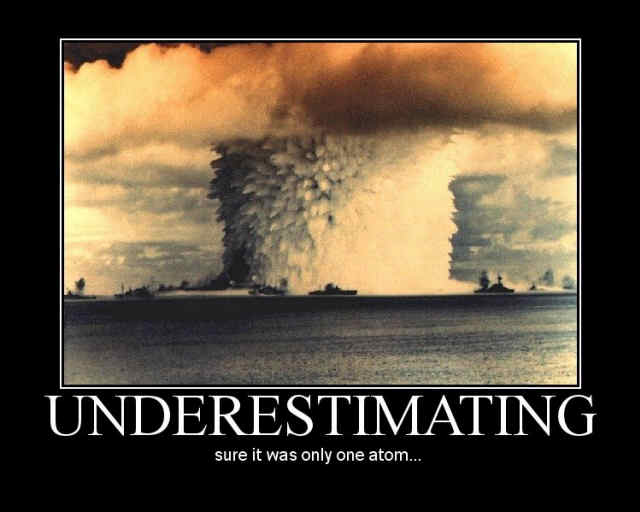

Underestimate the power and importance of
wee small things at your peril. The atom holds
extreme amounts of energy, and we depend on
the tiniest of prokaryotes for our survival on Earth.
It would be a mistake to underestimate the grit and power of some of nature’s smallest organisms. We could talk about this for months, but why don’t we stick to the discussion of prokaryotes and their ability to get along without conventional organelles that we began last week.

We can go farther in praise of the prokaryote by looking at how some of them manage to live in the most inhospitable environments; places that would kill us in seconds, or at least we hope they would. These are the “extremophiles;” the name makes them sound like Saturday morning cartoon superheroes.  For example, Thermococcus gammatolerans is the most radiation tolerant organism on Earth. It can laugh at gamma radiation levels 100x higher than other resistant organisms, even though it lives at the bottom of the sea.

As a result of the molecular biology revolution, many of the extremophiles are now called Archaea (Greek for “ancient”) or archaeabacteria, a completely group of organisms. Archaea are older than bacteria, and but they have some similarities to bacteria. Archaea are generally smaller than bacteria, but the cell wall of most archaea looks just like that of Gram+ bacteria. This is a thicker cell wall than that of Gram- bacteria, and takes up the Gram stain, hence the name Gram+.


The archaea cell wall is thick, and is contiguous with the cell membrane, 
like that of Gram+ bacteria. Gram- bacteria have thinner walls and 
they have a periplasmic space between the wall and the membrane.
Just looking at archaea and bacteria through a microscope makes it hard to tell the difference between these two distant relatives. It is at the molecular level that most of their differences become apparent. The way that archaea make RNAs is more eukaryotic than bacterial and while they both have cell walls, the lipids that make up archaeal membranes are quite different. Archaea lipids are hydrocarbon based, not fatty acid based like those of eukaryotes and bacteria. Also, archaeal cell walls lack the peptidoglycan that is characteristic of bacterial cell walls. Peptidoglycan synthesis is a common target of antibiotics, like penicillins, cephalosporines, and vancomycin.

This last difference might work out O.K. for us as humans. Not a single disease can be attributed to an archaea – yet. This is a big exception. Every other group of organisms on Earth has at least some members that can do humans harm, even if only inadvertently. Fungi, protozoa, bacteria, even plants can all cause us harm. One study says it is unlikely that we have just missed disease-causing archaea. About 0.38% of bacterial species cause disease, so if diversity in archaea is similar to that in bacteria, we should have found about 20 disease causing archaea by now.

Gum disease (periodontitis) has an outside chance of having an archaeal cause, but the evidence is sketchy. In a couple of studies, the presence of archaea in the mouth has correlated with gum disease; if archaea were present, then there was disease. Also, higher archaea number correlated to more severe disease. But archaea were only present in 1/3 of all cases of periodontitis – this is not good evidence to say archaea are the cause of periodontitis. This is the closest we have come to finding an archaeon with an anti-human bent.

Some archaea are thermophiles (heat loving); they don’t just like it hot, some require it really hot. Many thermophiles live in near undersea hydrothermal vents, where heat from the Earth’s mantle and core escapes into the ocean; basically ocean volcanoes.


The hydrothermal vent is an ecosystem that one
would be hard pressed to call home. Varies from 700˚C
to 4˚C, it is acidic, toxic, and radioactive. Yet many unique
prokaryotic and eukaryotic organisms live nowhere else.
To each his own.
Near a thermal vent, the temperature can reach 400-410˚C (700-720˚F) . The water doesn’t boil because of the great pressure exerted on it by all the water above it. No eukaryotic organism can survive at these temperatures, but thermophiles like T. gammatolerans do just fine. The hydrothermal vents pour out high levels of gamma type ionizing radiation from deep in the Earth, so it is handy that this archaeon is a multi-extremophile.

Only a few feet away from the vent the temperature of the ocean bottom will remain near freezing, about 4.5˚C. Other archaea (and some true bacteria) thrive in this cold environment. Called psychrophiles, cold tolerant archaea have cell walls that resist stiffening in water that is even below freezing temperature, and can fill there cytoplasm with anti-freeze proteins (AFPs; they create a difference between a solution melting point and its freezing point, called thermal hysteresis).

Between these two extreme environments, you can find quasi-conventional animals. As the hydrothermal vent water gives up its heat to the surrounding ocean, it creates an area that holds a temperature of about 10-15˚C. Many interesting animals have been found in this area, including the yeti crab and tube worms. Data from January 2012 describes a pure white octopus found at a depth of 2,394 meters. At this depth there is no light, so the octopus has no need for the elaborate camouflage mechanisms of color and texture. This octopod may represent a new species, but other white, vent-dwelling octopuses have been described previously, just not this far south.


This is the yeti crab (Kiwa hirsute). It is white because it lives
in the dark. It is furry because……..well, it makes the name
appropriate. Actually, the setae (hairs) contain bacteria that
may act to detoxify the water from the hydrothermal vents
where it lives. And it isn’t really a crab either, but I’m not
going to tell it so.
Ultimately, even these animals depend on the archaea for survival. No photosynthetic producers can survive at these depths, so the food chain starts with the chemosynthesizing prokaryotes, particularly those that use hydrogen sulfide to produce energy. Hydrogen sulfide is a major constituent in the hydrothermal vent output…. and would kill us quickly by binding to the enzymes in our mitochondria that perform ATP synthesis.

Some animals, like snails, eat the chemosynthesizing prokaryotes directly, while others predate the snails, etc. On the other hand, tube worms (Riftia pachyptila) get their energy directly from thermophilic proteobacterium that live inside the worm in a symbiotic relationship.

Other archaea live in high salt environments, like in the Dead Sea or the Great Salt Lake. They must be lonely, because given the high salinity, they are the only things living there (Water, Water Everywhere, But….). On the grosser end of the scale, some archaea thrive in human sewage plants, working well in environments without oxygen and high nitrogen contents.

Archaea have also been found in natural asphalt lakes, like near the La Brea region of Trinidad and Tobago. With toxic gases, high temperature, and practically no water at all, it was surprising that scientists found so many different kinds of prokaryotes, including several types of archaea. These 2010 findings suggest that life on other planets might not necessarily depend on water – that would be one heck of an exception!

But not all archaea are extremophiles, and they turn out to be much more common than we had thought. This isn’t just a numbers game, it turns out that we have been underestimating their effects on our lives all along. For instance, nitrogen fixation is crucial for crop production. A 2006 study by Schleper et al. in Norway suggests that there are many more ammonia oxidizing archaea in the soil than there are nitrogen fixing bacteria.


Archaea are responsible for much of the primary production that 
occurs in the soil and in the water. Just the methanogenic 
archaea alone are responsible for nearly 2% of all the carbohydrates
produced on Earth. Archaea contribute to the primary production 
of every ecosytem.
Further, current evidence suggests that archaea may represent 25-84% of all primary production (creation of carbohydrates and other organic compounds from inorganic carbon sources, whether by photosynthesis or chemosynthesis) in the upper layers of seawater. Primary production is the beginning of every food chain, so ultimately all of our food depends on archaea as well. To bad that we have been underestimating our dependence on these oldest of life forms. Who knows what our effects our life choices have been having on them all these years.

On the other hand, not all extremophiles are archaea either. Thermus aquaticus is a bacterium that lives in hot sulfur springs and geysers. It is a chemosynthesizing bacterium that has become important in molecular biology. Since its enzymes can tolerate high temperatures, it is useful for replicating DNA sequences in the lab using the polymerase chain reaction. One step in this reaction requires high temperature and would kill most other enzymes. 

Amazingly, this PCR technology and T. aquaticus polymerase has been crucial for helping us see how important the archaea have been in our evolution. In 1977, scientists Carl Woese and George Fox began DNA sequencing of some the extremophiles. They recognized that archaea were very different from eubacteria. The two groups must have diverged long long ago.


The three domains of life are shown here. The length of line shows 
the evolutionary distance between domains. You can see that 
Archaea are more like us than are the bacteria. You can’t tell from 
this chart, but Archaea are older too. They are the roots
of our family tree.
It turns out that Archaea are as closely related to eukaryotes as they are to eubacteria. This stood science on its ear. Up to this point, scientists had been arguing as to whether there were four, or five, or six kingdoms. Now they had to impose a higher classification which superseded all the kingdoms.

Woese’s evidence has led us define to the three domains of life. One domain is the eukaroytes, all the cells with a nucleus (with exceptions, but we can talk about those later), with linear chromosomes instead of one circular piece of DNA (again with exceptions), and with organelles. The second domain is the archaea and the third domain is the bacteria. Six kingdoms follow from these domains; archaea, bacteria, protista, fungi, plantae, and animalia.

Archaea, bacteria, and eukaryotes; we have shown that they are all different, and yet they all developed from some single precursor cell. Next time we will see if our discussion to this point gives us a roadmap to get from that ancient first cell to us.


Rogers, A., Tyler, P., Connelly, D., Copley, J., James, R., Larter, R., Linse, K., Mills, R., Garabato, A., Pancost, R., Pearce, D., Polunin, N., German, C., Shank, T., Boersch-Supan, P., Alker, B., Aquilina, A., Bennett, S., Clarke, A., Dinley, R., Graham, A., Green, D., Hawkes, J., Hepburn, L., Hilario, A., Huvenne, V., Marsh, L., Ramirez-Llodra, E., Reid, W., Roterman, C., Sweeting, C., Thatje, S., & Zwirglmaier, K. (2012). The Discovery of New Deep-Sea Hydrothermal Vent Communities in the Southern Ocean and Implications for Biogeography PLoS Biology, 10 (1) DOI: 10.1371/journal.pbio.1001234

For more information and classroom activities on archaea, hydrothermal vents, chemosynthesis, and domain/kingdoms, see:

Archaea and extremophile bacteria –

Hydrothermal vents –

Chemosynthesis –

Domains/kingdoms -

Wednesday, July 30, 2014

Does Life Come In XXXS?

Biology concepts – characteristics of life, archaea, bacteria, mycoplasma, synthetic biology, symbiosis, parasitism, nanobacteria, genome

As part of this blog, we have talked about some pretty small life. Wolffia globosa is the smallest flowering plant, only 0.6 mm long. We also talked about archaea, a different kingdom than bacteria, but still on the smallish side of life. The tardigrade is the toughest animal, but is also one of the smallest, at 100 µm (0.00394 inch).


The organism on the top is T. dieteri, and arthropod, just
as is any crab or spider. The size is deceiving. The pictures
on the bottom are to scale and are copepods, also
arthropods. The organism on the top is a parasite of the
organisms on the bottom. The small blue line? That would
be the scaled size of T. dieteri. So…. it’s SMALL.
The question for today is – is there a minimum size for life? Candidates might include bacteria or archaea; heck there’s an arthropod, Tantulacus dieteri, that's only 85 µm long! As long as we can keep finding smaller and smaller cells, we know that the minimum size for life is that small or smaller. So we keep looking – you’d be surprised how important it is to keep looking for smaller life.

Here’s one thing we should be able to agree on, viruses don’t get to play in our game. Viruses are very small, but they're not life! We’ve talked about this before - the seven characteristics of life (see this post). Viruses need a host in order to replicate, they don’t manage homeostasis, and they aren’t cells, so they aren’t life.

So how small has actual life become? Let’s assume that since tardigrades and T. dieteri are over 50 µm, huge when compared to some bacteria, our current minimum for life is probably a bacterium or archaea.

Let’s go straight to the genus of smallest bacteria we know about – the mycoplasma (from mykes = fungus, and plasma = formed). They were first described in 1898, but the observer didn’t have a clue what he was looking at; hence the fungal part of the name.

Mycoplasma don’t have the traditional cell wall of many bacteria, so they look different and this might be why they were mistaken for fungi. Whatever the scientists thought of them, they were confusing enough to be roundly ignored for 50 years. Rediscovered in the 1950’s-1960’s, this time they were thought to be L-forms of bacteria. L-forms are organisms that for some reason have lost their cell wall.

There are stable forms of L-bacteria; they can live divide and live on without their cell wall. There are also unstable L-forms as well; those that may revert to walled bacteria at any moment. Are mycoplasma simply bacteria that have lost a cell wall? Nope. They didn’t have a cell wall to lose. They have no cell wall genes, so if they had a cell wall, it was millions of years ago, before they became their own genus.


The difference between some free living cells. You can
probably see the E. coli in bright green, but you may have
to squint to see the mycoplasma above it. It’s pink. Really,
it’s there. Compare these sizes to those of the arthropods
above. 1 mm is equal to 1000 µm.
Mycoplasma is really, really, SMALLLLLLL.
Mycoplasma are generally described in the range of 0.2-0.8 µm in diameter. But this is a little misleading, because they are often not spherical. Even without a cell wall, they can take interesting three-dimensional forms and maintain them. Mycoplasma pneumoniae, which causes a form of …..….. anyone?…….. right, pneumonia, is pear shaped, so its 0.25 µm diameter is actually the measurement on its short side.

So mycoplasma are small, but they still have to play by the rules. They contain DNA and salts and proteins and ribosomes and other things that take up room. A single ribosome is about 50 nm in diameter (0.05 µm or 0.00000005 m), so there must be a certain volume required for the cell to function – a minimum size for life.

Which of the mycoplasma species is the smallest? Mycoplasma genitalium is considered to be the smallest mycoplasma known, and the smallest form of free-living organism - my gosh – you can fit about 400 M. pneumoniae inside one E. coli! As such, it is the current minimum size for life that we have. M. genitalium is 200 nm (0.2 µm) x 600 nm (0.6 µm), so they’re pretty dawg on small. Let’s put it this way, there are 25,400,000 nm in one inch – mucho small.

It is important to note that M. genitalium is free living, but does need some help. It uses cholesterol in its membrane but doesn’t make it itself. It picks it up from the cells that it lives near……wait for it….. your genital epithelium.


One of the human diseases that is becoming more
convincingly associated with M. genitalium is pelvic
inflammatory disease (PID). Resulting when many
different sexually transmitted diseases go untreated,
PID can cause permanent damage to the reproductive
organs of women. It is important to get treatment early.
The inflammation of PID may be associated with the
fallopian tubes or ovary, and will cause a chronic pain
in the lower abdomen, bleeding and pain on urination.
M. genitalium is a cause of non-gonococcal urethritis (inflammation of the urethra). A late 2013 review states that 1-3% of the general population is infected with M. genitalium, more than with gonorrhea. It is linked to pelvic inflammatory disease, and the review cites studies showing that people infected with this mycoplasma are more at risk for HIV and have more dual infections. It’s a sexually transmitted organism, just another reason for proper restraint. But even though it's helped out by your genital epithelium, it can live on its own and divide outside a host, so it's considered a free-living organism.

The idea of free-living is important because M. genitalium also has a very small genome (amount of DNA in one cell, including the list of all its genes). M. genitalium has about 580 kbp of DNA where kbp = kilobase pairs. Remember that DNA is doubled stranded (usually) so each base is paired with another. Knowing this, we count them as a unit. In all, M. genitalium has just 520 or so genes; it can make about that many proteins.

Genome size could be another way of determining the minimum size of life - what's the minimum number of genes or number of base pairs of DNA for an organism to still meet all seven characteristics of life? As of summer 2014, no organism smaller in size than M. genitalium has been described, but there have been some other organisms discovered with smaller genomes.

Nanoarchaeum equitans was thought to have the smallest gene for a while, with only 491 kbp of DNA. It is an archaea that lives on the edge of hydrothermal vents at the bottom of the ocean. But it is an obligate symbiont with another archaea; it can’t survive without its partner, so can you say it has the minimal genome? It relies on another organism’s DNA.


On the left is the leafhopper in which N. deltocepahlinicola makes
his home. Well inside its cells that is. The leafhopper survives on
phloem and xylem; high in carbs but little protein. The bacterium
makes the amino acids the leafhopper can’t in exchange for energy
in the form of ATP. On the right is a colored photomicrograph of
the abdomen. The red is one type of endosymbiont bacteria,
the green is N. deltocephalinicola.
This is also true of Carsonella ruddii (159 kbp, 182 genes) and Nasuia deltocephalinicola. They are bacteria that must live inside insect cells, like those of grasshoppers. N. deltocephalinicola has the smallest known genome (112 kbp, 137 genes), but it doesn’t even make ATP, it steals it from the arthropod cells. This could hardly be considered free living, and so it can’t be considered the minimal genome for life. And even at that, their cell sizes are still bigger than M. genitalium.

So why is it important to find the minimal size and minimal genome for life? So we can use the information. J. Craig Venter (of the human genome project) wanted to develop a synthetic form of life (synthetic biology); a bacterium that could be developed to provide hydrogen for energy or eat waste to reduce pollution. Others say we need to know so that we can better recognize life on other planets, or life that may have come here from other planets (astrobiology).

Being J. Craig Venter, develop a synthetic form of life is exactly what he and his research institute did. It’s interesting that Venter was one of the scientists that first sequenced the entire M. genitalium genome in 1995. Some 15 years later, Venter’s JCVI-syn1.0 (2010) was the first synthetic life, housing 1000 kbp and 500 or so genes. The genome was based on that of another mycoplasma, M. mycoides. They modified the genome, and introduced it into a cell membrane that had been evacuated of all its constituents. The resulting cell was capable of growing, dividing, you know…. living.

If M. genitalium represents our current estimate for the minimum size of life, it’s only because we’re thinking of life as we know it. Perhaps we have already found life that is smaller, and the minimum size is actually much smaller than M. genitalium.


This is a photomicrograph of a meteorite from Mars. The
small spheres (like the ones the arrows point to, are
supposedly nanobacteria. Proof of life on Mars,
contamination from Earth nanobacteria, or just mineral
spheres that look a little like incredibly tiny bacteria?
The answer is C.
Something termed a nanobe and something else called a nanobacterium were described 20-30 years ago. Nanobes were first found in the rocks that came up during oil drilling in Australia, while nanobacteria were also found in surface rocks.  The size of both (about 1/20 size of M. genitalium) negates their use of ribosomes and DNA. They stain for DNA, but this may be artifact, the artificial result of other things picking up the stain.

But nanobes/nanobacteria have their proponents. Some scientists say that since no DNA has been exhibited, they are a completely different form of life, so size restriction (big enough to hold ribosomes) doesn’t apply. Nanobacteria are also claimed to be important in human disease, as these structures are found in many calcifications of diseased tissues.

On the other hand, nanobacteria are probably just mineral formations. A 2013 study showed that they form spontaneously from many different biological fluid samples, and their appearance in diseased tissues is more a sign of disease than a cause of it. We’ll just have to keep looking for something smaller.

Next week, another question tackled and dissected - think pink.


Manhart LE (2013). Mycoplasma genitalium: An emergent sexually transmitted disease? Infectious disease clinics of North America, 27 (4), 779-92 PMID: 24275270

Wu CY, Young L, Young D, Martel J, & Young JD (2013). Bions: a family of biomimetic mineralo-organic complexes derived from biological fluids. PloS one, 8 (9) PMID: 24086546

Gibson DG, Glass JI, Lartigue C, Noskov VN, Chuang RY, Algire MA, Benders GA, Montague MG, Ma L, Moodie MM, Merryman C, Vashee S, Krishnakumar R, Assad-Garcia N, Andrews-Pfannkoch C, Denisova EA, Young L, Qi ZQ, Segall-Shapiro TH, Calvey CH, Parmar PP, Hutchison CA 3rd, Smith HO, & Venter JC (2010). Creation of a bacterial cell controlled by a chemically synthesized genome. Science (New York, N.Y.), 329 (5987), 52-6 PMID: 20488990


Wednesday, August 14, 2013

How Prokaryotes Shape Up

Biology concepts – morphology, eubacteria, coccus, rod, bacillus, spirilla, spirochete, prosthecae, halotolerant,

The standard menu of prokaryotic shapes may seem boring, but they
really perform specific functions. The underlined types are those we
hear about most often, perhaps you’ve heard of “bacillus” more often
called “rod.” Bacillus is actually the name of one genus of rod-shaped
bacteria, but the name has taken over in many circles, like Kleenex for
facial tissue. Most likely, all the forms evolved from rods, and you can
see how this might be possible from the cartoon.
Most prokaryotes are small - really small. At best they show up as small dots in most microscopes. But we know they have definite shapes. The question is, is the shape of a bacterium an important detail?

Question of the Day – What shapes can prokaryotes take and are their shapes important?

We are taught in school that bacteria have three shapes; spheres, rods, and spirals, but there are actually many more. The second part of our question is just as important to think about. Does it matter what shape a bacterium takes? Do you really think it’s random… really? After all the things we have discussed in this blog?

A 2007 paper summarized the evidence for why scientists believe the morphology (shape) of bacteria must be important. Here's a brief summary. One - genera of bacteria will always show the same subset of shapes. Why spend the energy to maintain the genetic blueprint if it isn’t important? We have talked before about how genes that are not necessary are allowed to drift, but morphology genes don’t drift.

Two - changes in environmental conditions or pressures will bring changes in the morphology of many prokaryotes. What is more, the same change will be bring the same morphology alteration again and again. This implies both regulation and specific functions for different shapes. Shape must be important if it is worthy of controlled regulation.

And three - archaea and eubacteria are very different, as different as you and a pine tree, but they tend to fall within the same types of morphologies. Each representative morphology must be adaptive (important for survival and propagation) if they turn up again and again in very unrelated organisms.

This and other papers by Dr. Young also discuss the ways morphology confer advantages. Our morphology discussion should also include size. Small bacteria usually have shorter generation times. This is important because faster developing bacteria usually out-compete larger ones for limited resources and nutrients. Therefore, most bacteria are very small.

The typical classroom answer for small size is that bacteria do not have intracellular transport systems, so all movement of nutrients and important molecules must be by diffusion. A big cell means too slow a transit time and death. Also, being small is a good way to maximize surface area to volume, so lots of room is given for possibly contacting food, while keeping diffusion time fast.

These are valid reasons to be small, but size is just as important in reducing the chances of being eaten. There probably hundreds of thousands of different single-celled eukaryotes that feed exclusively or mostly on bacteria. But bacteria are cannibals as well. They'll eat other bacteria, and if times are bad enough, may feed on their own kind.

To avoid being the midnight snack of some protozoan, bacteria have several choices. You can be small and fast, or you might opt to become huge - too large to ingest or even be recognizes as food.

There is a lot going on in this collage. On the left is the aggregates that
cocci can form based on the plane in which they divide. The only form
possible for rods is the strepto- form since they always divide through
their short side. The pictures on the right are to illustrate the size
difference between E. coli and T. nambibiensis. If E.coli were the size of
a tic tac candy, the crater seen below could not hold T. nambibiensis.
Bacteria can also join forces by attaching to one another and become too large to consume, either in long chains, large masses, or complex biofilms. Would you pull out a fork and spoon and try to choke down an elephant in the wild – what about a herd of elephants all stuck together?

But back to shape - what might different shapes do for different bacteria? Let’s look at some amazing prokaryotic shapes in terms of several factors: nutrient acquisition; predators; cell division; attachment; dispersal; motility; and differentiation of function.

Coccus
Cocci (the plural of coccus, from the Greek kokkus = berry) are round bacteria. Spherical is a safe shape since it gives the maximum surface area for a given volume. However, spherical doesn’t necessarily mean small. Thiomargarita namibiensis is a spherical bacterium, but it is the second largest prokaryote we know of. If an E. coli cell was the size of a tic tac, T. namibiensis would have a diameter a bit larger than the Barringer Meteor crater in Arizona (see picture above).

Rod
The rod is probably the oldest prokaryotic morphology. It is most probable that cocci were short rods that kept getting shorter, and that other shapes we will talk about are also modifications of rods. So give the rods (of which E. coli is one) the respect they deserve – it may seem mundane later when we talk about weird shapes, but the rod is the mother of all shapes.

Rods show that motility comes into play as a reason for shape. The rod shape, longer than wide, is the fastest mover in response to chemical signals (chemotaxis, chemo = chemical and taxis = arrangement), the chemical trail left by a potential meal for example. Becoming longer and thinner is also a good way to increase apparent size (reduce predation) and provide more surface area (for food collection).

Spirals
Spiral shaped bacteria are faster through viscous fluids – so this shape is probably an adaptation to allow movement in different fluids. Many spiral bacteria live in environments thicker than water, so moving faster than predators would be important.

The spiral shape of spirilla is usually thicker and flatter than that of spirochetes, and another difference is that spirochetes often have different attachments for their flagella (whip-like oars for movement).

Predation may also play a different role in spiral shape development. Arthrospira platensis (a cyanobacteria) grows as a spiral.  It also known as spirulina, a potential superfood, but promoters usually say it’s a blue-green algae, not a bacterium.

Spirulina is eaten by a protist that can turn left on its long axis up to six times to ingest the A. platensis. Low and behold - A. platensis can reverse it spiral direction in the face of predation so that the ciliate would have to spin right to eat it, and it can’t do that. 

On the left is Caulobacter crescentus, a crescent shaped prokaryote. It
takes two forms, a swarming crescent with flagella for times of
plenty and a stalked crescent for times when food is scarce. In some
parts of the image, you can see the stalk. Atopobium rimae is on the
right, a coccobacillus. A. rimae is a constituent of normal oral flora,
but can cause periodontal disease as well. Its looks remind me of a
microscopic Easter egg hunt.
Coccobacilli

I call these bacteria elliptical; one nice example is Atopobium rimae. They look like footballs, not quite a rod (bacilli are one genus of rod shaped bacteria), and not yet reduced to a sphere.

Many coccobacilli are pathogenic, including the organisms that cause chancroid STDs, brucellosis, pneumonia, infectious blindness, bacterial flu, and whooping cough. However, a link between shape and disease causation escapes me. I haven’t found any evidence that someone has even asked the question. Maybe you can.

Crescent shaped
These are rod shaped bacteria that have become curved. Vibrio bacteria are an important group of crescents (named because they looked like they vibrated as they moved)….. oh, and because they cause a lot of disease.

The crescent shape can be important for movement. Vibrio alginolyticus swims forward just fine, but when it encounters a flat surface – an area where food might be gathered – it swims backward. Its crescent shape keeps it bumping into the flat surface. This keeps it longer in the area of food. In some crescents, a mutation making them straight means that they lose their motility and ability to find food, so it must be important.

Triangular and square
Yes, there are prokaryotes that look very much like triangles or squares. I am listing them together because the thing they have most in come is that they are usually halophiles (halo = salt, and philic = loving) or are halotolerant.
Both of the bacteria above are halophilic, meaning they love salt.
Haloarcula japonica (left) and Holoquadratum walsbyi (right)
for perfectly shaped to form contiguous sheets of cells. This
helps them float parallel to the surface of the water they live in.
These are archaeal extremophiles, but it is interesting that
archaea and bacteria use the same sets of shapes.

The triangular example I have for you is Haloarcula japonica, so named because it was discovered in a Japanese salt evaporation field. Another member of the same genus is H. quadratum, which you might guess, is square.

But it isn’t just this genus that take on definite geometric shapes, there is another salt-loving arachaean called Haloquadratum walsbyi that is also square. Mind you, these are not cubiodal bacteria, and the H. japonica is not a triangular prism. They are very flat; H. walsbyi, for example, is 5 µm square but only 0.1 µm thick. The same can be said for the triangular H. japonica.

They all tend to grow as flat masses, like little floating mosaics. Their flat shapes keep them buoyant and floating parallel to the water’s surface. Their geometry then provides the largest surface toward the sun, in order to pick up the most heat and energy. Sounds logical to me.

Star
These are my favorites, the stars of the bacterial world. The far left is
Stella vacuolata. Those bubbles in the middle are gas – yes, bacteria get
gas too. The middle photomicrograph is Prosthecomicrobium.
Prostheacae are the arms, and are right in the name, even though these
are very short prosthecae. On the right is Ancalomicrobium adetum. Its
prosthecae are much longer and are used to deter predators, amongst
other things. There is even one very long rod bacterium that has a
star cross section.
It must take quite a bit of regulation to build and maintain a star shape. The ones I have seen come in a couple of varieties. Some are definitely star-shaped because of their cell body. Stella vacuolata is star-shaped, as are Prosthecomicrobium species. The reason for a star-shaped adaptation--- I don’t know - patriotism maybe?

There are also bacteria that have projections from their cell body that make them look like fireworks as they explode; see the picture of Ancalomicrobium adetum. The projections are called prosthecae (Greek for appendage), and can serve many functions. They can increase surface area without increasing mass for better diffusion. They can make a bacterium large enough to not be eaten. They may also catch more water, to help non-motile bacteria be moved along by the current.

Other
We have only touched the surface of the morphologies that prokaryotes can assume. There are others that look like Y’s (bifid bacteria), some that look like connected lollipops, some that look like segmented worms, and at least one that builds a net of connected tubules near black smokers at the bottom of the ocean (Pyrodictum abyssi).

Among the many other shapes possible for bacteria, the bifids are
interesting (left). Certain proteins are produced only in the ends of
the bacteria, and if they need more of that protein, especially for
attachment, one way to get it is to have more ends. On the
right is an archaea found at the bottom of the ocean. The fine lines
are actually tubular projections with fine nets of bacteria cell
inside. The cell bodies are the nodular areas seen nonsymmetrically.
Just how do prokaryotes construct and control their shape? This is an active area of research and may be important for medicine. We have seen that shape affects function and survival, so new antibiotics might just work to turn rods into cocci or stars into blobs.

A 2011 paper shows that zinc metal is essential for proper morphology. Zinc is an important part of several proteins that control which DNA is read to make proteins (zinc finger transcription factors), so this is evidence that discrete controls are in place to define a bacterium’s morphology.

A 2013 study shows that E. coli rod shape is determined mostly by its cell wall. If the cell wall was removed, it took 4-6 generations for the rod shape to be recovered. If mutations in certain lipoproteins or penicillin binding proteins were present, the bacteria progeny would always remain spherical. These genes are not even used in producing the cell wall, so it is apparent that many genes are needed just to maintain cell shape. My current research concerns the ability to bend rod bacteria into tiny balloon animals.

Next week we will start our series of exceptions and core concepts for the year. We begin by looking at the elements of life - there's more than you think, and then we'll look at the four types of biomolecules. 


Ranjit DK, & Young KD (2013). The Rcs stress response and accessory envelope proteins are required for de novo generation of cell shape in Escherichia coli. Journal of bacteriology, 195 (11), 2452-62 PMID: 23543719

Bayle L, Chimalapati S, Schoehn G, Brown J, Vernet T, & Durmort C (2011). Zinc uptake by Streptococcus pneumoniae depends on both AdcA and AdcAII and is essential for normal bacterial morphology and virulence. Molecular microbiology, 82 (4), 904-16 PMID: 22023106  

Young KD (2007). Bacterial morphology: why have different shapes? Current opinion in microbiology, 10 (6), 596-600 PMID: 17981076


Wednesday, May 15, 2013

Biodiversity Counts!

Biology concepts – biodiversity, kingdoms of life, animalia, plantae, fungi, protist, archaea, bacteria, extant, extinct

It seems that contests counting things in jars
always involve food. M&Ms, jellybeans, candies,
and gumballs are common things to estimate. I like
another estimate game – how many grains of sand
can be held in one human hand?
Amazingly, only about 10,000.
At some point in our lives, we have all tried to win the prize by guessing how many jellybeans are in the jar. Number estimation is a skill few people possess, just try ordering mulch by the square yard – you end up with either half as much as you need or buried in pine bark!

There is a large group of scientists playing the jellybean game for a living, but their jar is the entire earth. The jellybeans are species of organisms. The prize? Well, you don’t get to keep the jellybeans; and just how will we know who wins?

The Question of the Day – How many species of life are there on Earth?

It seems like a straight forward question, but let’s look at it in a bit more detail before we try to answer it.  What is it we're counting, animals? Animals and plants? Let’s include everything – everything that is considered alive. So what is considered alive? You can have a great discussion as to what should be considered alive, but for these purposes, let’s stick to the kingdoms of life as taught in every biology class – Archaea, Bacteria, Protists, Fungi, Plants, and Animals.

So now that we know the biodiversity we are assessing, we need to define our unit. We said above that we would count species, but it isn’t that simple. We have discussed before what constitutes a species in general – those animals that can breed and produce fertile offspring. So we don’t count ligers and tigons (crosses between tigers and lions), but should we count all the hybridizations of orchids? More than 24,000 species are named already, with about 800 added each year. For our purposes, and for most people estimating species, we will stick with those found in nature, unaided by man.

In 2012, Japanese botanists crossed two orchids and
created a new orchid. No big deal right, orchids are
crossed all the time. But this was the first time that a
photosynthesizing orchid was crossed with a purely
parasitic orchid that doesn’t perform photosynthesis.
They think the plants are doing some photosynthesis –
they’re pretty anyway. Is this a species we should
count in our estimate?
We are counting species, but is it live (extant) species or all species including those that are extinct? The World Wildlife Federation estimates that between 0.01% and 0.1% of all species go extinct each year. However many species we end up with as an estimate for all life on Earth, this is a huge number of species to lose EVERY YEAR. It is scary to think how many undiscovered species will go extinct this year. There must be thousands and thousands of species that we will never get to describe using a live specimen or discover how they might add to the diversity of the planet.

Scientists refer to “catalogued” species, but does that refer to those alive at the time the were described, or any species that has ever been described and named? Scientists estimate that extant species account for only 3% of everything that has lived on Earth, so including all species would greatly increased the overall number. However, in most estimates of species on Earth, the species being talked about are alive now, except for the one went extinct just this second, and the one that will go extinct two minutes from now, and the one….


The opposite action is also occurring; we discover new species every year. Most people think that perhaps a few new species are found each year, but it’s really in the thousands. The International Institute for Species Exploration (IISE) at Arizona State University publishes a list each year of the newly discovered species. For 2011 the list was more than 18,000 species long! You can see from the picture (below, right) that many discoveries were made in every kingdom of life in the decade of the 2000’s, more than 170,000 discoveries in all.

This graph shows the relative number of new species in many groups of
organisms. The scales are variable for each group and can’t be compared
amongst the groups. The trends show that in some groups, more and
more species are being discovered each year, while in others, fewer and
fewer are being found. In still others, some years a bunch are found and
some years provide few or no new species.
If 170,000 were discovered in ten years, the total number of species on Earth must be huge. Let’s break down the numbers of catalogued species and the estimates by kingdom.

Animalia – These are what most people think of when asked to name a species. Most animals are relatively big and can be seen in everyday life. The number of catalogued species includes the dinosaurs and humans, birds and sponges.  The numbers in each group vary greatly.

People love to study birds – so we have probably found a greater percentage of the total number of birds than we have worms. We have named about 10,000 species of birds, and more than 22,000 species of annelids (segmented worms). But there are probably many more annelids that we have not found as compared to birds. Therefore the estimate for annelids will be harder to make and perhaps less accurate.

For all animals, the total number of catalogued species by 2010 was 1.2 million. A recent paper (2011, Mora et al.) has predicted the number of species in most kingdoms based on several mathematical models. The authors predict that there are more than 9.9 million animal species on the Earth and in the oceans. According to this estimate, we have found only 12% of all the animals on Earth!

Meet the Giant Gippsland Earthworm 
(Megascolides australis). It can reach 3meters 
(10 feet) in length. First described in 1878, this 
worm lives in the deep clay soil in a small 
area in Australia. Similar worms live in North
America, but they are rarely observed.
Plantae – Plants include the non-vascular mosses, the vascular, spore-forming ferns and horsetails, the seed bearing gymnosperms (conifers and such) and the fruiting and flowering angiosperms. So many of these organisms are crucial for human life (medicine, food, oxygen!) we have done a good job of cataloguing them. The 2011 Mora paper surmises that we have already described nearly 50% of the total number of species.

Their methodology uses a lot of math to relate the number of higher taxa (phylums, orders, families) that are known in well-described kingdoms to the possible number in lesser studied groups. They found that there was a consistent pattern in which the more families there are now can be used to predict how many total genera there might be, and each level then can be used to predict the number in kingdoms for which the number of higher taxa are known. They also use methods to predict unknown higher taxa, so they can be included in the species estimate.

For plants, the Mora group predicts that there are 314,000 species extant on Earth. Other estimates also come out at about 300,000 species, but they include algae, which are actually protists, not plants.

Fungi – These organisms range from the invisible to the visible. In fact, in the paleoworld, fungi represented the largest organisms on Earth. Even today, the largest single organism is a fungus in the Malheur National Forest in Oregon, where a single 8,000 year old honey mushroom covers 2,200 acres (8,900 square meters) of land.

On the top is a fossilized prototaxite fungus in Saudi Arabia 
that once reached 20 ft (6 m) in height and was the tallest 
organism on the early Earth (this one is on its side). On the 
bottom is the national forest where a single honey mushroom mat 
has killed 2200 acres of trees. Each mushroom is a clone,
connected by a rhizoid mat just under the ground.
If you compare plant diversity to fungi, fungi win hands down. Described species of fungus lag behind; only 43,000 species have been named, but there is much more room to add new species. Estimates are that by the time we are done, whenever that is, we will have nearly 700,000 different mushrooms and other fungi.

Protista – Protists are a bit of a catch-all kingdom. Some have aspects that make them look like plants; they perform photosynthesis or they have central vacuoles. Others are much more animal-like. Overall, they are free-living organisms that are usually single celled or made up of many cells not forming tissues. Two examples show you the diversity in this group. Giant sea kelp (Macrocystis pyrifera) is a type of brown algae that can grow at a rate of 2 ft/day and can reach a length of 300 ft (91.5 m), while picoplankton are 0.2 microns (0.00000002 m) in diameter, are single celled, and may or may not perform photosynthesis.

The estimates for the number of protists vary greatly. A 1998 study indicated that they had no reason to believe the total number of protist species would be greater than 3000. However, a 2005 report puts the estimate number anywhere from 140,000 to 1.6 million. The Mora group’s paper estimates that about 73,000 will be found; that’s 57,000 more than have been described as of 2010.

Archaea and Bacteria – These are the prokaryotes. They live in tar pits and arm pits; they own the planet and probably outer space as well. There are more bacteria in a scoop full of dirt then people who have ever lived on Earth. But for the purposes of our discussion today, the important part is that same scoop of dirt has thousands of undiscovered bacteria.

The J. Craig Venter Institute is leading the Global Ocean Sampling Expeditions to discover new marine microorganisms. A pilot expedition in 2003 identifed 1800 new species in just a couple of months. This was followed by global expeditions in 2005-2009 and an expedition to the European waterways in 2009-2010. They will analyzing the data and naming species for decades to come.

This map represents the expeditions of the Global Ocean Sampling Projects
of the Venter Institute. As they traveled, they would acquire 200-400 gallon
samples of water from different depths every 200 miles and put them
through a series of filters to catch smaller and smaller organisms. The filters
would be dried and used for DNA analysis. In just a couple of months, 1.2
million genes were identified using this methodology.
Most bacteria and archaea can’t be grown in the lab because we don’t know what they require to live. This makes them hard to describe and classify.  Therefore, the Venter Institute uses DNA techniques from dried whole organisms to identify new “species.”

But perhaps classification is not the proper term for these organisms. I talked to Dr. Mora about why prokaryotes were not analyzed to the same degree as other kingdoms in their paper.  He rightfully pointed out that species definitions don’t really apply to prokaryotes as neatly as they do other types of organisms.

Certainly they don’t conform to the mating and fertile offspring definition of species since they don’t mate. Also, since they swap DNA as usual business (lateral gene transfer), who is to say where one species stops and another begins.

Other sources are little more daring in projecting possible numbers of prokaryotes. It is possible that there are a billion distinct bacteria and archaea, but it more likely that the number is in the 10-20 million range.

What are our final numbers when add up all the estimates? Predicted species numbers for life on Earth range from 11.3 million in the Mora paper, to perhaps more than 1 billion if you include prokaryotes. If we leave the bacteria out of the equation, there could still be as many as 30,000,000 forms of life on the planet. We have described about 2 million (according to IISE), so only 28,000,000 left to find!


Mora, C., Tittensor, D., Adl, S., Simpson, A., & Worm, B. (2011). How Many Species Are There on Earth and in the Ocean? PLoS Biology, 9 (8) DOI: 10.1371/journal.pbio.1001127
 
ADL, S., SIMPSON, A., FARMER, M., ANDERSEN, R., ANDERSON, O., BARTA, J., BOWSER, S., BRUGEROLLE, G., FENSOME, R., FREDERICQ, S., JAMES, T., KARPOV, S., KUGRENS, P., KRUG, J., LANE, C., LEWIS, L., LODGE, J., LYNN, D., MANN, D., MCCOURT, R., MENDOZA, L., MOESTRUP, O., MOZLEY-STANDRIDGE, S., NERAD, T., SHEARER, C., SMIRNOV, A., SPIEGEL, F., & TAYLOR, M. (2005). The New Higher Level Classification of Eukaryotes with Emphasis on the Taxonomy of Protists The Journal of Eukaryotic Microbiology, 52 (5), 399-451 DOI: 10.1111/j.1550-7408.2005.00053.x